Cardiac output is the volume of blood the heart pumps per minute and a central concept in cardiovascular physiology. Boards emphasize its equation, the determinants of stroke volume — preload, afterload, and contractility — and the Frank-Starling relationship that links them.
check_circleAfterload is the resistance the ventricle pumps against.
The Cardiac Output Equation
Cardiac output (CO) is the product of heart rate (HR) and stroke volume (SV): CO = HR × SV. A typical resting value is about 5 liters per minute. Because it is the flow that supplies the entire body, cardiac output must rise to meet increased demand, as in exercise, by increasing heart rate, stroke volume, or both. The cardiac index normalizes output to body surface area.
Determinants of Stroke Volume
Stroke volume, the blood ejected per beat, is set by three factors. Preload is the degree of ventricular filling before contraction (end-diastolic volume), influenced by venous return and blood volume. Contractility (inotropy) is the intrinsic strength of contraction, increased by sympathetic stimulation and drugs like digoxin. Afterload is the resistance the ventricle must overcome to eject blood, largely determined by arterial pressure and vascular resistance.
The Frank-Starling Relationship
The Frank-Starling law states that, within limits, the more the ventricle is filled (greater preload), the more forcefully it contracts and the greater the stroke volume. This occurs because stretching cardiac muscle optimizes the overlap of contractile filaments. It allows the heart to automatically match its output to venous return and to balance the outputs of the two ventricles on a beat-to-beat basis.
Regulation and Clinical Relevance
The body regulates cardiac output through the autonomic nervous system (sympathetic activity raises heart rate and contractility; parasympathetic activity lowers heart rate) and through changes in preload and afterload. These principles underpin clinical management: fluids increase preload, inotropes increase contractility, and vasodilators reduce afterload, and heart failure is understood as the failure of cardiac output to meet the body's demands.
Try it now
3 real questions on Cardiac Output
Straight from the Progress Note question bank, each with a cited explanation. No account needed.
123
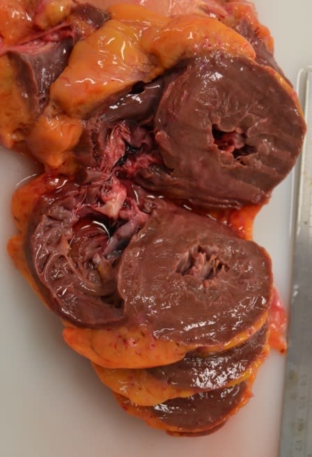
A 70-year-old man with a history of aortic stenosis undergoes cardiac catheterization. Simultaneous pressure measurements reveal a peak systolic left ventricular pressure of 210 mmHg and a peak systolic aortic pressure of 120 mmHg. Echocardiography shows a left ventricular ejection fraction of 65% with concentric left ventricular hypertrophy and a wall thickness-to-radius ratio greater than normal. The following is seen upon autopsy. Which of the following best explains why ejection fraction is preserved despite the chronic pressure overload?
Mikael Häggström, M.D., CC0, via Wikimedia Commons
Answer: B. According to the Law of Laplace, wall stress (σ) = (pressure × radius) / (2 × wall thickness). In chronic pressure overload (e.g., aortic stenosis), the ventricle responds with concentric hypertrophy — parallel addition of sarcomeres increases wall thickness without increasing chamber radius — which normalizes systolic wall stress and preserves ejection fraction. This adaptation maintains systolic function but at the cost of impaired diastolic compliance, leading to diastolic dysfunction over time. Eccentric hypertrophy (option A), characterized by sarcomeres added in series, is the response to volume overload (e.g., aortic regurgitation, dilated cardiomyopathy) and increases chamber radius. Preload enhancement via Frank-Starling (option C) does not explain preserved EF in the setting of chronic outflow obstruction.
Source: Otto, C. M., et al. (2021). 2020 ACC/AHA guideline for the management of patients with valvular heart disease. Journal of the American College of Cardiology, 77(4), 450–500.
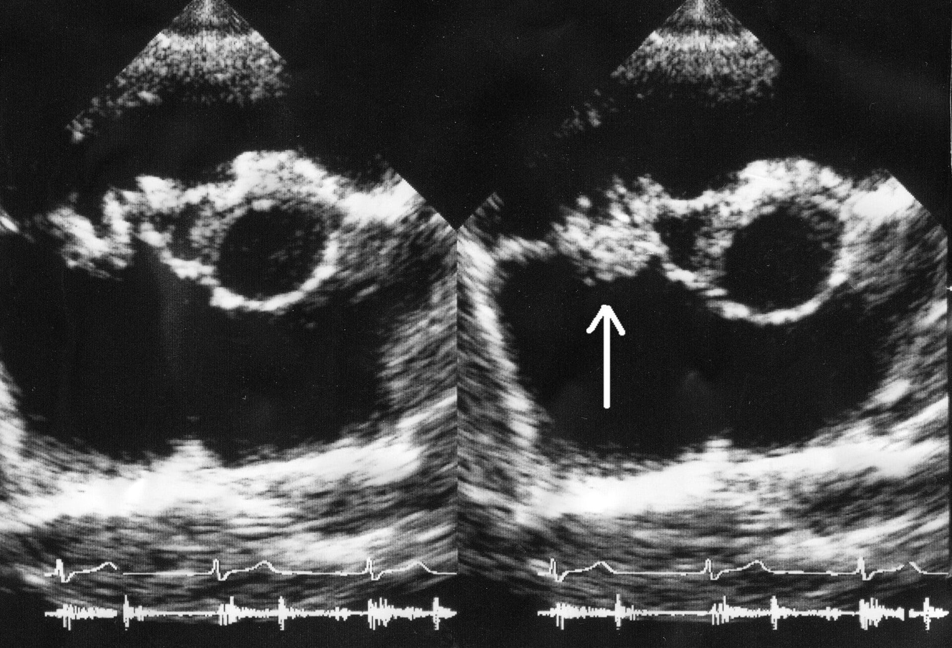
A 42-year-old man with a history of intravenous drug use presents with fever, new cardiac murmur (regurgitant), and microhematuria. Blood cultures grow Staphylococcus aureus. An echocardiograph is shown below. Which of the following best explains the pathogenesis of bacterial endocarditis and the relationship between valvular vegetation formation and septic embolization in this patient?
Daisuke Koya, Kazuyuki Shibuya, Ryuichi Kikkawa and Masakazu Haneda. derivative work: Materialscientist, CC BY 2.0 <https://creativecommons.org/licenses/by/2.0>, via Wikimedia Commons
Answer: B. Acute bacterial endocarditis results when circulating bacteria seed valve endocardium, typically at sites of endothelial injury caused by turbulent flow, prior valve disease, or traumatic seeding from IVDU. Bacteria adhere to valve endocardium via adhesins (for S. aureus: fibronectin-binding proteins, collagen adhesins) or to preexisting platelet-fibrin deposits from minor valve trauma. Once adherent, bacteria replicate rapidly within a protected nidus of platelets, fibrin, and bacteria—the vegetation. This nidus shields bacteria from antibiotics (poor penetration) and from immune cells (limited access). Septic emboli form when vegetation fragments detach and embolize to distant organs, causing septic infarcts and abscesses. The friable, poorly organized nature of acute bacterial vegetations makes them prone to embolization, unlike the more organized vegetations of subacute endocarditis. Options A, C, D, and E invoke incorrect or incomplete mechanisms.
Source: Murdoch, D. R., et al. (2009). Clinical presentation, etiology, and outcome of infective endocarditis in the 21st century. Archives of Internal Medicine, 169(5).
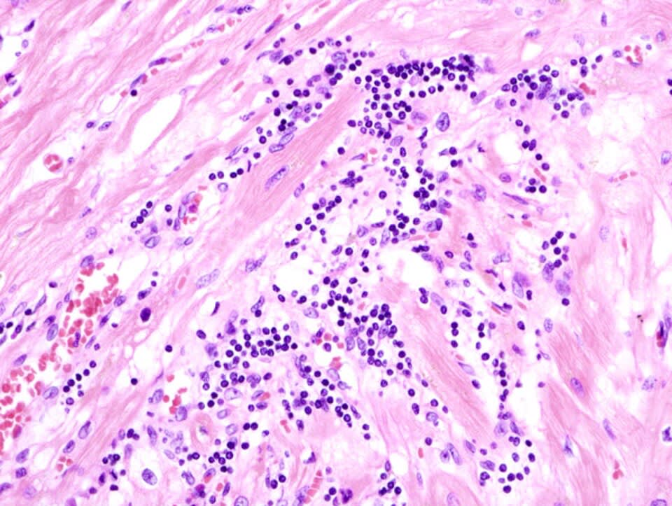
A 32-year-old woman presents with acute-onset chest pain, dyspnea, and palpitations. ECG reveals diffuse ST-segment elevation (except in aVR and V1) with PR-segment depression. Cardiac biomarkers are elevated (troponin I 2.8 ng/mL). Transthoracic echocardiography shows global hypokinesis with preserved ejection fraction and a small pericardial effusion. Endomyocardial biopsy is shown below. Which of the following best explains the pathophysiology and the relationship between viral infection, myocardial inflammation, and the characteristic clinical and pathologic findings observed in this patient?
KGH, CC BY-SA 3.0 <https://creativecommons.org/licenses/by-sa/3.0>, via Wikimedia Commons
Answer: B. Acute myocarditis results from viral infection of myocardial myocytes (commonly enterovirus, adenovirus, parvovirus B19, influenza, or coronavirus); viral replication triggers both innate and adaptive immune responses. Innate immunity activates pattern recognition receptors (TLRs) on myocytes and macrophages, triggering release of pro-inflammatory cytokines (IL-6, TNF-alpha, IL-1) and type I interferons. The adaptive immune response follows: dendritic cells and macrophages process viral antigens and present them via MHC-I (for CD8+ T cells) and MHC-II (for CD4+ T cells). CD8+ cytotoxic T lymphocytes recognize virus-infected myocytes and destroy them via perforin-mediated pore formation and granzyme-induced apoptosis, as well as via Fas-FasL interactions. The myocardial infiltrate reflects this T-cell-mediated destruction: CD8+ T cells predominate, accompanied by CD4+ T cells and macrophages. Myocyte necrosis releases cardiac troponins into circulation, producing troponin elevation. Diffuse subepicardial inflammation causes diffuse ST-segment elevation and PR-segment depression (pericardial involvement). The relatively preserved ejection fraction in early myocarditis reflects global rather than focal dysfunction; severe cases may progress to fulminant myocarditis with cardiogenic shock. Option A incorrectly emphasizes direct viral lysis over immune-mediated destruction; option C overemphasizes autoimmunity without viral involvement; options D and E invoke incorrect mechanisms (complement-mediated lysis, TNF-alpha-only mechanism).
Source: Caforio, A. L., et al. (2013). Current state of knowledge on aetiology, diagnosis, management, and therapy of myocarditis: A position statement of the European Society of Cardiology Working Group on Myocardial and Pericardial Diseases. European Heart Journal, 34(33).
Your next questions are ready.
The question bank picks up from here: it adapts to what you just missed and brings it back right before you'd forget it.
Cardiac output equals heart rate multiplied by stroke volume (CO = HR × SV). A typical resting cardiac output is about 5 liters per minute, and it increases during exercise by raising heart rate, stroke volume, or both.
What determines stroke volume?expand_more
Stroke volume is determined by three factors: preload (ventricular filling before contraction), contractility (the intrinsic strength of contraction), and afterload (the resistance the ventricle pumps against). Increasing preload or contractility raises stroke volume, while increasing afterload lowers it.
What is the Frank-Starling law?expand_more
The Frank-Starling law states that, within physiologic limits, greater ventricular filling (preload) leads to a more forceful contraction and a larger stroke volume. This lets the heart automatically match its output to venous return and balance the two ventricles.
What is the difference between preload and afterload?expand_more
Preload is the amount the ventricle is stretched by filling before it contracts (end-diastolic volume), reflecting venous return and blood volume. Afterload is the resistance the ventricle must overcome to eject blood, mainly determined by arterial pressure and systemic vascular resistance.
Free weekly high-yield email
One high-yield concept + one question, every week
Study smarter with a short weekly email. No spam — unsubscribe anytime.
✓ You're in — talk soon.
Reading is one thing. Remembering is another.
Turn this guide into questions you’ll actually remember
Progress Note turns concepts like this into adaptive questions and spaced review, so difficult topics come back to you exactly when you’re about to forget them.