Hypokalemia is a serum potassium below 3.5 mEq/L and is one of the most common electrolyte disturbances. Because potassium governs cardiac and neuromuscular excitability, boards emphasize its causes, the characteristic ECG changes, and correct repletion including magnesium.
Evidence-based · For exam prep, not medical advice
update Updated October 2026
Date of last content change
Built by surgery and anesthesiology residents·Written for USMLE Step 1
bolt The 60-second version
check_circleHypokalemia is a serum potassium below 3.5 mEq/L.
check_circleCauses fall into GI loss, renal loss, and transcellular shift into cells.
check_circleECG shows flattened T waves, ST depression, and prominent U waves.
check_circleSymptoms include muscle weakness, cramps, and arrhythmias.
check_circleCoexisting hypomagnesemia must be corrected or potassium will not normalize.
Causes
Hypokalemia arises through three mechanisms. Gastrointestinal loss comes from vomiting, diarrhea, or nasogastric suction. Renal loss results from diuretics (loop and thiazide), hyperaldosteronism, and some tubular disorders. Transcellular shift moves potassium into cells with insulin, beta-2 agonists, and alkalosis. Poor intake alone rarely causes significant hypokalemia unless combined with losses.
Clinical Features and ECG
Mild hypokalemia is often asymptomatic. As it worsens, patients develop muscle weakness, cramps, fatigue, constipation from ileus, and, importantly, cardiac arrhythmias. The classic ECG evolution is flattening of the T wave, ST-segment depression, and the appearance of prominent U waves, with increased risk of ventricular arrhythmias, especially in patients on digoxin.
Diagnosis
Evaluation combines the history (vomiting, diarrhea, diuretics) with assessment of acid-base status and, when the cause is unclear, urine potassium and blood pressure to separate renal from extrarenal losses. Low urine potassium suggests extrarenal (GI) loss or transcellular shift, whereas inappropriately high urine potassium points to renal wasting, such as hyperaldosteronism.
Treatment
Treatment corrects the deficit and the underlying cause. Oral potassium is preferred for mild to moderate hypokalemia, with cautious intravenous replacement for severe or symptomatic cases or when the patient cannot take oral therapy. Crucially, coexisting hypomagnesemia must be corrected, because low magnesium promotes renal potassium wasting and makes hypokalemia refractory to replacement.
Try it now
3 real questions on Hypokalemia
Straight from the Progress Note question bank, each with a cited explanation. No account needed.
123
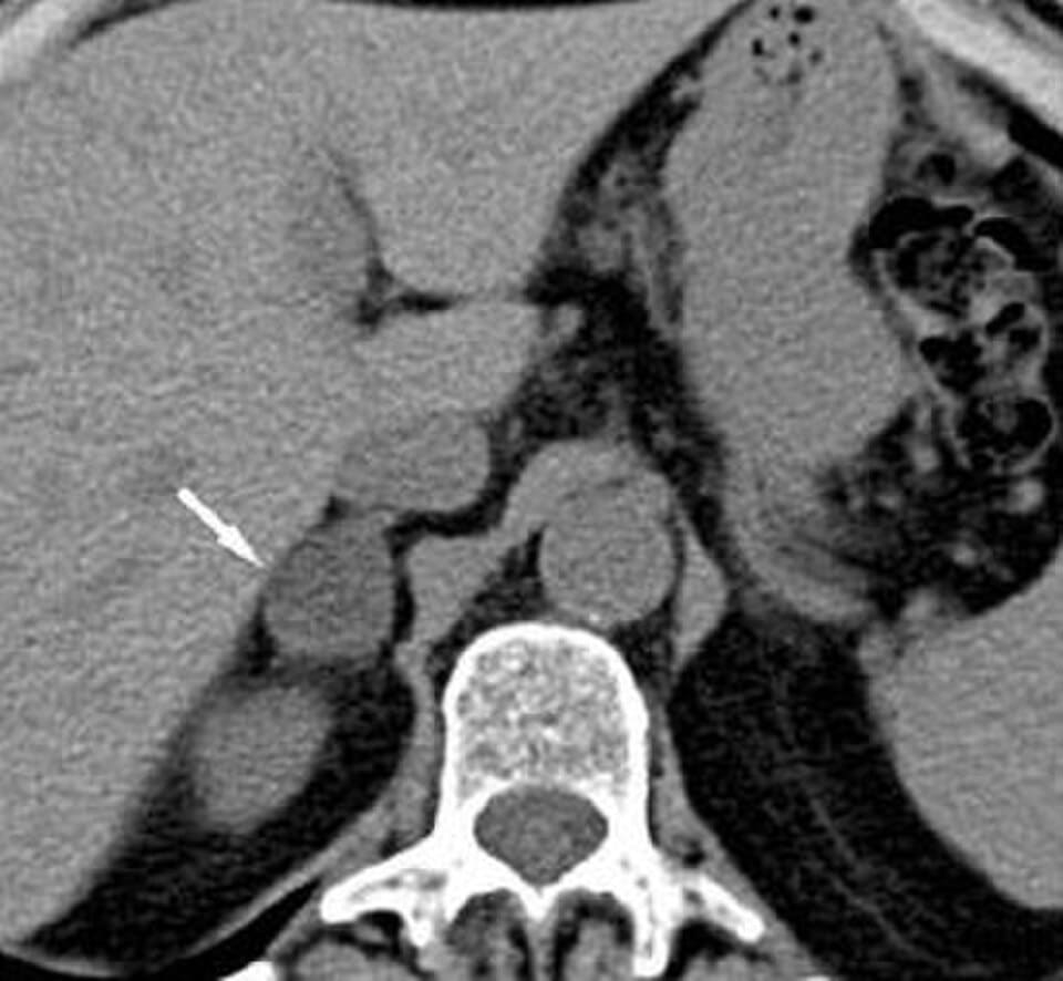
A 48-year-old woman with resistant hypertension on three antihypertensive agents is found to have spontaneous hypokalemia (potassium 3.0 mEq/L). Plasma aldosterone-to-renin ratio is markedly elevated. CT of the abdomen is shown below. The suppressed plasma renin activity observed in this patient is best explained by which of the following mechanisms?
Antonino Guerrisi, Daniele Marin, Mahbubeh Baski, Pietro Guerrisi, Federica Capozza, and Carlo Catalano, CC BY 4.0 <https://creativecommons.org/licenses/by/4.0>, via Wikimedia Commons
Answer: A. In primary hyperaldosteronism due to an aldosterone-producing adenoma, autonomous aldosterone secretion causes renal sodium and water retention, expanding extracellular fluid volume and increasing renal perfusion pressure; this volume expansion suppresses renin release from the juxtaglomerular apparatus through normal negative feedback, independent of the classic renin-angiotensin-aldosterone cascade that would otherwise drive aldosterone secretion. This combination of high aldosterone with suppressed renin is the key biochemical signature distinguishing primary from secondary hyperaldosteronism, the latter of which, as in renal artery stenosis, would show elevated renin. ACE inhibitors increase, rather than decrease, renin release by interrupting angiotensin II-mediated negative feedback, the opposite of what is described in one distractor option.
Source: Vaidya, A., et al. (2018). The expanding spectrum of primary aldosteronism: Implications for diagnosis, pathogenesis, and treatment. Endocrine Reviews, 39(6).
From our NCLEX bank
A nurse receives a prescription reading potassium chloride 20 mEq intravenous push for a client with hypokalemia. Which action by the nurse is correct?
Answer: A. Potassium chloride is never administered by intravenous push, because a rapid rise in serum potassium causes fatal cardiac arrest, so the nurse questions the prescription (option A). Giving it slowly over 5 minutes while watching the monitor (B) is still a push and remains lethal. Administering it because the potassium is low (C) confuses the indication with a safe route. Diluting it in 10 mL and pushing slowly (D) is also a push rather than the required infusion. Potassium is always diluted in a large volume and infused at a controlled rate through a pump.
Source: U.S. Food and Drug Administration. (2023). Potassium chloride injection [Drug label]. U.S. Food and Drug Administration.
From our NCLEX bank
A client has intravenous potassium chloride ordered for hypokalemia. Which action by the nurse is essential for safe administration?
Answer: D. Intravenous potassium is never given by push; it is diluted and infused on a pump at a controlled rate with monitoring, so this action is essential for safety (option D). Giving potassium by IV push (A) can cause fatal cardiac arrest. Administering it undiluted (B) is dangerous and causes severe vein irritation and dysrhythmias. Increasing the rate to finish quickly (C) raises the risk of lethal hyperkalemia and cardiac effects. Controlled, diluted infusion with cardiac and renal monitoring protects the client. The nurse checks the potassium level, renal function, and urine output and assesses the site for irritation.
Source: U.S. Food and Drug Administration. (2023). Potassium chloride injection [Drug label]. U.S. Food and Drug Administration.
Your next questions are ready.
The question bank picks up from here: it adapts to what you just missed and brings it back right before you'd forget it.
Hypokalemia results from gastrointestinal loss (vomiting, diarrhea), renal loss (diuretics, hyperaldosteronism), or a shift of potassium into cells (from insulin, beta-agonists, or alkalosis). Diuretic use and GI losses are among the most common causes.
What are the ECG changes in hypokalemia?expand_more
Hypokalemia classically causes flattened T waves, ST-segment depression, and prominent U waves, along with an increased risk of ventricular arrhythmias. These changes are especially dangerous in patients taking digoxin.
Why do you give magnesium with potassium?expand_more
Low magnesium promotes renal potassium wasting and makes hypokalemia resistant to potassium replacement. Correcting a coexisting hypomagnesemia is therefore essential, or the potassium level will not normalize despite supplementation.
How is hypokalemia treated?expand_more
Treatment replaces potassium — orally for mild to moderate cases and intravenously (cautiously) for severe or symptomatic hypokalemia — while correcting the underlying cause and any coexisting magnesium deficiency.
Free weekly high-yield email
One high-yield concept + one question, every week
Study smarter with a short weekly email. No spam — unsubscribe anytime.
✓ You're in — talk soon.
Reading is one thing. Remembering is another.
Turn this guide into questions you’ll actually remember
Progress Note turns concepts like this into adaptive questions and spaced review, so difficult topics come back to you exactly when you’re about to forget them.