How nephrotic syndrome is tested on Step 1
Expect a child with periorbital edema after an infection, or an adult with edema and a systemic disease, plus a biopsy description.
Complications of protein loss
Each complication reflects a lost plasma protein.
Primary podocytopathies
Minimal change disease and FSGS both injure podocytes but differ in prognosis.
Causes compared
The patient and the biopsy findings point to the cause.
| Disease | Light microscopy | EM / IF | Associations |
|---|---|---|---|
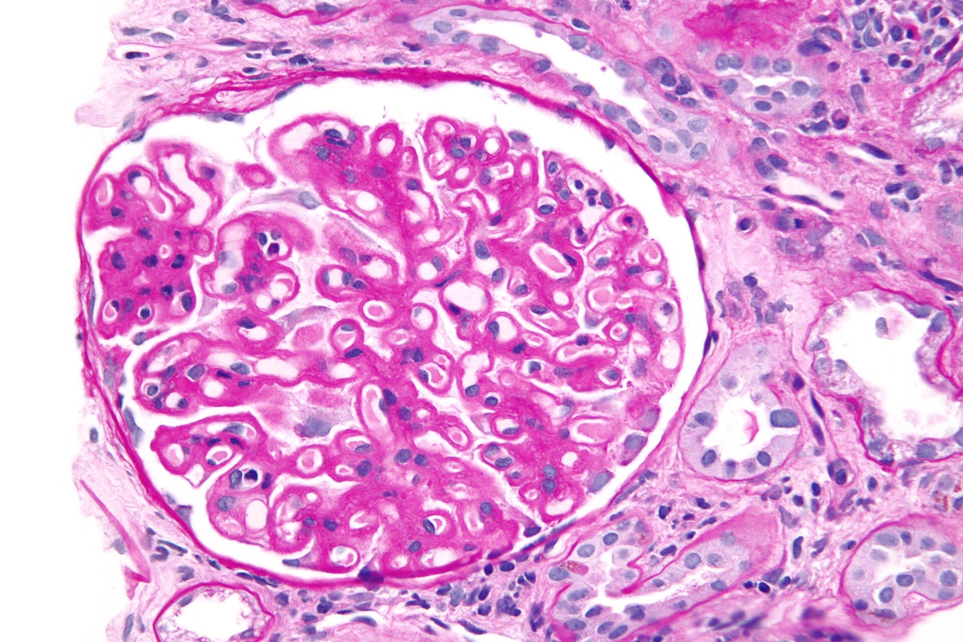
| Minimal change | Normal | Effaced foot processes; IF negative | Children; Hodgkin lymphoma; NSAIDs |
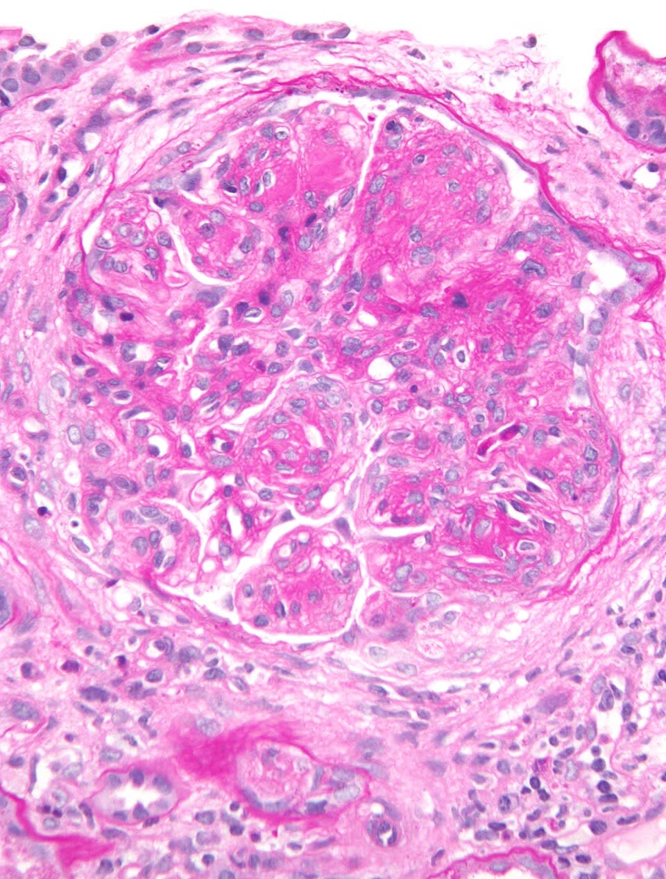
| FSGS | Segmental sclerosis and hyalinosis | Effacement; IF usually negative | HIV, heroin, sickle cell, obesity, APOL1 |
| Membranous | Diffuse GBM thickening | Subepithelial deposits, "spike and dome"; granular IgG | Anti-PLA2R; lupus (class V), hepatitis B, solid tumors, NSAIDs, penicillamine |
| Diabetic nephropathy | Mesangial expansion; Kimmelstiel-Wilson nodules | GBM thickening | Long-standing diabetes |
| Amyloidosis | Congo red-positive deposits | Apple-green birefringence | Plasma-cell dyscrasia, chronic inflammation |
Hyperfiltration in early diabetes is followed by albuminuria; ACE inhibitors or ARBs and SGLT2 inhibitors slow progression.
Treatment and a worked example
Treat the cause, reduce proteinuria, and manage complications.
- Glucocorticoids for minimal change disease (most children remit).
- ACE inhibitor or ARB to reduce proteinuria; loop diuretics for edema; statins for hyperlipidemia.
- Immunosuppression for selected FSGS and membranous nephropathy.
- Anticoagulation when thrombosis occurs (and sometimes prophylactically in high-risk membranous disease).
A 58-year-old has edema and 6 g/day proteinuria. Biopsy shows diffuse GBM thickening, subepithelial deposits, and silver-stain "spikes." Serum anti-PLA2R antibody is positive. He develops sudden flank pain and hematuria. What happened?
- GBM thickening with subepithelial deposits and spikes = membranous nephropathy.
- Anti-PLA2R positivity indicates primary (autoimmune) disease.
- Membranous nephropathy carries a high risk of thrombosis from urinary loss of antithrombin III.
- Sudden flank pain and hematuria suggest renal vein thrombosis.
Answer. Renal vein thrombosis, a hypercoagulable complication of membranous nephropathy.