Chronic kidney disease (CKD) is kidney damage or reduced function lasting more than 3 months. Step 1 focuses less on staging trivia and more on physiology: why failing kidneys cause anemia, bone disease, acidosis, hyperkalemia, and bleeding, and which drugs slow progression.
Evidence-based · For exam prep, not medical advice
update Updated October 2026
Date of last content change
Built by surgery and anesthesiology residents
Written for USMLE Step 1 · Free to read
bolt The 60-second version
check_circleCKD = GFR < 60 mL/min/1.73 m² or markers of damage (such as albuminuria ≥ 30 mg/g) for more than 3 months.
check_circleDiabetes and hypertension are the leading causes; cardiovascular disease is the leading cause of death.
check_circleAnemia: low erythropoietin → normocytic anemia; treat iron deficiency first, then an erythropoiesis-stimulating agent.
check_circleCKD-mineral bone disorder: phosphate retention and low calcitriol → hypocalcemia → secondary hyperparathyroidism → renal osteodystrophy.
check_circleUremia: pericarditis, encephalopathy (asterixis), pruritus, nausea, and platelet dysfunction (treat bleeding with desmopressin).
check_circleACE inhibitors or ARBs and SGLT2 inhibitors slow progression in albuminuric CKD; dialysis or transplant for kidney failure.
target What you'll be able to do · aligned to USMLE Step 1 · Renal
RENALDefine CKD and classify it by GFR and albuminuria.
PHYSExplain the pathway from reduced GFR to secondary hyperparathyroidism.
PATHRelate anemia, acidosis, hyperkalemia, and uremic bleeding to lost kidney functions.
PHARMIdentify therapies that slow CKD progression and treat its complications.
How CKD is tested on Step 1
Expect a patient on dialysis or with long-standing diabetes and questions about lab patterns (calcium, phosphate, PTH, hemoglobin) and the mechanism of each complication.
GFR categories (mL/min/1.73 m²). KDIGO also stages albuminuria: A1 < 30, A2 30–300, A3 > 300 mg/g.
Causes and progression
Whatever the cause, loss of nephrons leads surviving nephrons to hyperfilter, which injures them in turn.
Uremic symptoms usually appear only when GFR is very low.
Slowing progression and a worked example
Treatment targets blood pressure, albuminuria, and complications, then kidney replacement.
ACE inhibitor or ARB for albuminuric CKD (expect a small creatinine rise; stop if it rises sharply).
SGLT2 inhibitors slow CKD progression in patients with and without diabetes.
Avoid nephrotoxins: NSAIDs, unnecessary contrast; adjust renally cleared drug doses.
Kidney replacement: hemodialysis, peritoneal dialysis, or transplantation (best long-term survival).
functions Worked example — lab pattern
A patient on hemodialysis has bone pain. Labs: calcium 8.0 mg/dL, phosphate 7.2 mg/dL, PTH markedly elevated, alkaline phosphatase elevated. Explain the pattern.
Low GFR retains phosphate, and low calcitriol reduces calcium absorption.
Low calcium and high phosphate stimulate the parathyroids.
PTH rises (secondary hyperparathyroidism) and drives bone resorption.
High bone turnover raises alkaline phosphatase and causes renal osteodystrophy (osteitis fibrosa cystica).
Answer. Secondary hyperparathyroidism from CKD-mineral bone disorder, causing renal osteodystrophy.
Straight from the Progress Note question bank, each with a cited explanation. No account needed.
123
A 38-year-old man presents with new-onset hypertension, flank pain, and hematuria. His father died of a ruptured intracranial aneurysm and had a history of 'kidney problems.' CT of the abdomen shows bilaterally enlarged kidneys, and the gross specimen image below is representative of the expected pathology. Genetic testing identifies a mutation in PKD1. Which of the following best explains the mechanism of renal cyst formation in this patient's disease?
CDC/Dr. Edwin P. Ewing, Jr., Public domain, via Wikimedia Commons
Answer: A. Autosomal dominant polycystic kidney disease, most commonly caused by mutations in PKD1 (encoding polycystin-1), disrupts the mechanosensory function of the primary cilium in renal tubular epithelial cells. Loss of normal ciliary calcium signaling raises intracellular cAMP, which drives both epithelial proliferation and fluid secretion into the tubular lumen, progressively expanding fluid-filled cysts that replace normal parenchyma. ADPKD is also associated with intracranial berry aneurysms, explaining this patient's family history of stroke. Autosomal recessive polycystic kidney disease involves a distinct fibrocystin mutation and presents in infancy, while VHL disease causes renal cysts and tumors through an unrelated tumor suppressor mechanism.
Source: Torres, V. E., et al. (2007). Autosomal dominant polycystic kidney disease. The Lancet, 369(9569).
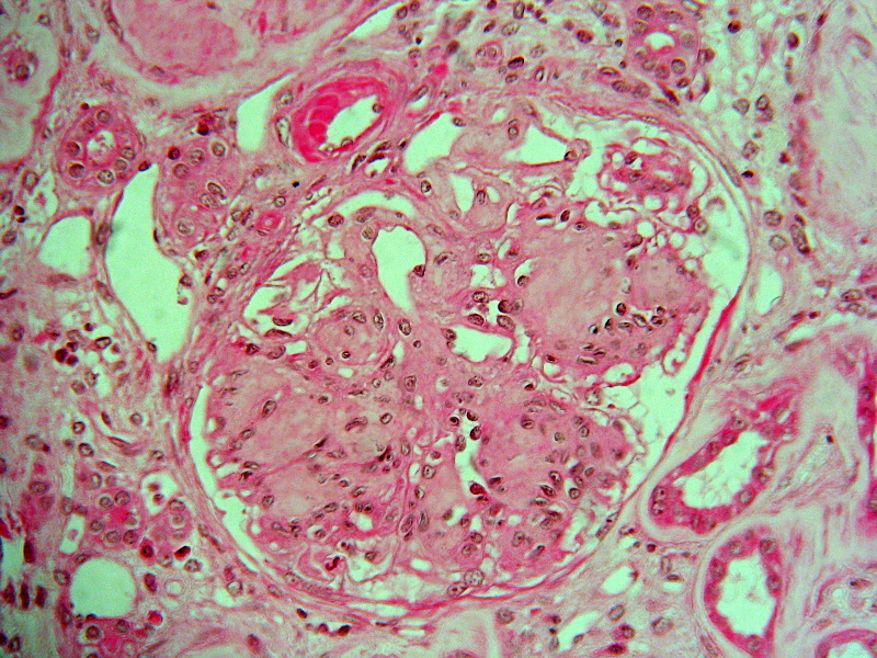
A kidney biopsy shows nodular mesangial expansion with thickened glomerular basement membranes on light microscopy. Which underlying condition is most consistent with these histologic findings?
Doc.mari, CC BY-SA 3.0 <https://creativecommons.org/licenses/by-sa/3.0>, via Wikimedia Commons
Answer: B. Diabetic nephropathy is characterized by nodular mesangial expansion (Kimmelstiel-Wilson nodules) and thickened glomerular basement membranes. Hypertensive nephrosclerosis typically shows hyaline arteriolosclerosis rather than nodules. Minimal change disease lacks significant light microscopy findings. Poststreptococcal and IgA nephropathies show immune complex deposition with hypercellularity. These nodular lesions are classic for diabetic kidney disease.
Source: Tervaert, et al. (2010). Pathologic classification of diabetic nephropathy. Journal of the American Society of Nephrology.
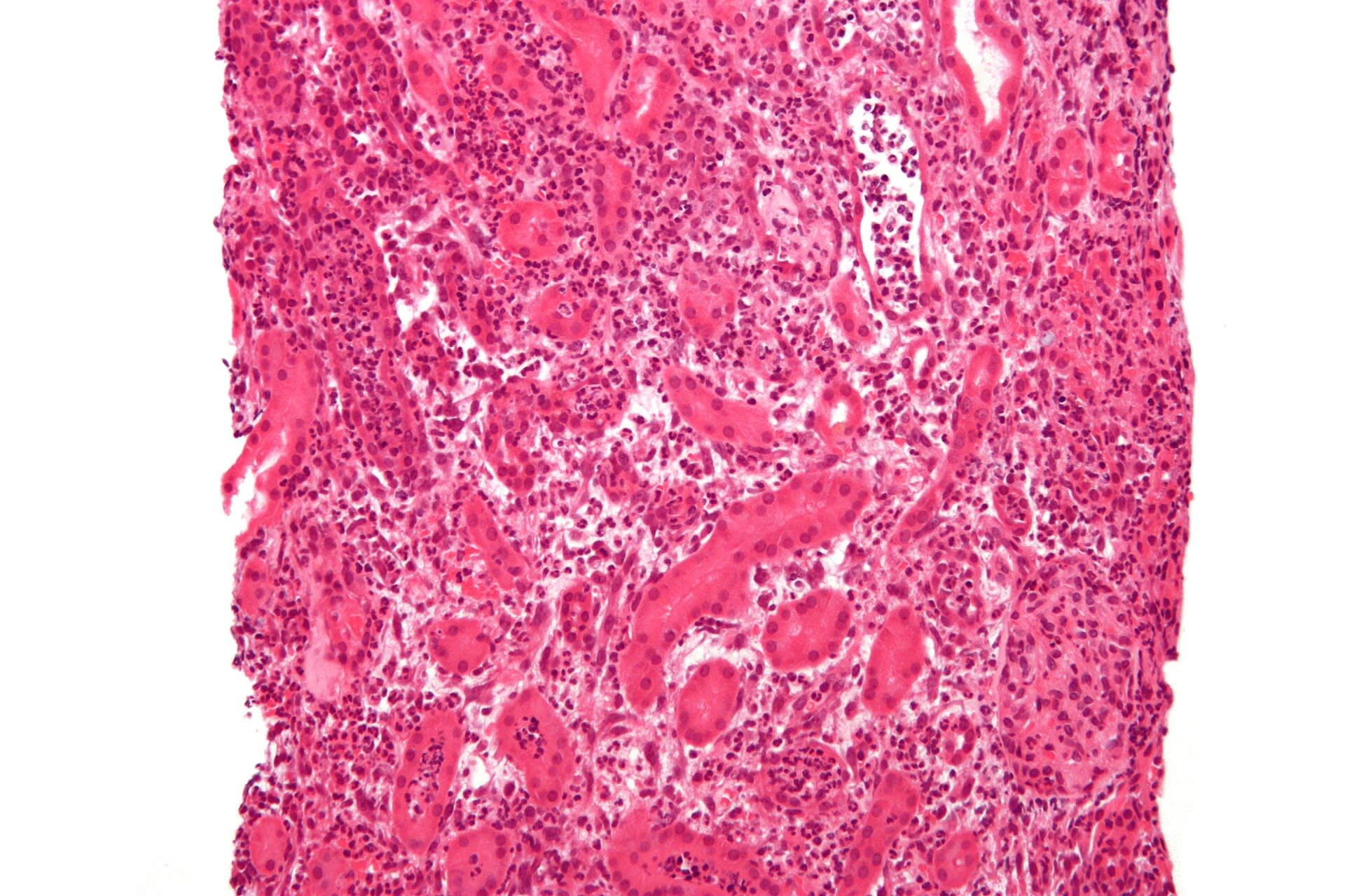
A 28-year-old woman presents with fever, flank pain, and dysuria. Urinalysis shows leukocyte esterase positivity and nitrites. Gross examination of the kidney would most likely reveal which finding?
Nephron, CC BY-SA 3.0 <https://creativecommons.org/licenses/by-sa/3.0>, via Wikimedia Commons
Answer: B. Acute pyelonephritis is a bacterial infection of the kidney, most commonly caused by ascending gram-negative organisms such as E. coli. Gross pathology typically shows patchy, yellow-white abscesses and streaking of the renal medulla due to neutrophilic infiltration along the tubules. Option A describes chronic pyelonephritis or chronic renal disease with scarring. Option C is more consistent with amyloidosis or other infiltrative diseases. Option D represents renal infarction, which lacks suppurative inflammation. Option E describes polycystic kidney disease.
Peritubular interstitial cells make less erythropoietin. Iron deficiency and shorter red cell survival contribute. The anemia is usually normocytic.
Why is PTH high in CKD?expand_more
Phosphate retention and low calcitriol lower serum calcium, and high phosphate and FGF23 add to the stimulus. The result is secondary hyperparathyroidism.
How is uremic bleeding treated?expand_more
Desmopressin (which releases von Willebrand factor) and dialysis. The defect is platelet dysfunction, not a low platelet count.
What is the leading cause of death in CKD?expand_more
Study smarter with a short weekly email. No spam — unsubscribe anytime.
✓ You're in — talk soon.
Reading is one thing. Remembering is another.
Turn this guide into questions you’ll actually remember
Progress Note turns concepts like this into adaptive questions and spaced review, so difficult topics come back to you exactly when you’re about to forget them.