Glomerulonephritis (Nephritic Syndrome): The USMLE Step 1 Guide
Glomerulonephritis is inflammation of the glomeruli that damages the filtration barrier and lets red cells into the urine. Step 1 tests the nephritic pattern, the timing of infection-related disease, immunofluorescence patterns, crescents, and which diseases lower complement.
Evidence-based · For exam prep, not medical advice
update Updated October 2026
Date of last content change
Built by surgery and anesthesiology residents
Written for USMLE Step 1 · Free to read
bolt The 60-second version
check_circleNephritic syndrome: hematuria with RBC casts and dysmorphic RBCs, hypertension, oliguria, azotemia, and proteinuria below the nephrotic range.
check_circlePoststreptococcal GN: 1–3 weeks after pharyngitis or 3–6 weeks after skin infection; low C3; subepithelial humps; granular ("lumpy-bumpy") immunofluorescence.
check_circleIgA nephropathy: hematuria within days of a mucosal infection; mesangial IgA deposits; normal complement; most common GN worldwide.
check_circleRapidly progressive GN = crescents (fibrin, parietal epithelial cells, macrophages); classified by immunofluorescence: linear, granular, or pauci-immune.
check_circleAnti-GBM (Goodpasture): antibodies to type IV collagen (α3 chain) → linear IgG; with lung hemorrhage it is Goodpasture syndrome.
check_circleLow complement: poststreptococcal, lupus, membranoproliferative, cryoglobulinemic GN. Normal complement: IgA, anti-GBM, ANCA vasculitis.
target What you'll be able to do · aligned to USMLE Step 1 · Renal
PATHDistinguish nephritic from nephrotic syndrome.
PATHDifferentiate poststreptococcal GN from IgA nephropathy by timing, complement, and pathology.
IMMUNOClassify RPGN by immunofluorescence and associated antibodies.
RENALIdentify which glomerular diseases lower serum complement.
How glomerulonephritis is tested on Step 1
Vignettes give a timeline after infection, an immunofluorescence or EM description, or a pulmonary-renal syndrome, and ask for the diagnosis or mechanism.
Nephritic vs nephrotic. Some diseases (membranoproliferative GN, lupus nephritis) can show either pattern.
Infection-related GN vs IgA nephropathy
The interval between infection and hematuria separates the two.
Timing. IgA nephropathy flares during or right after the infection (synpharyngitic); PSGN follows a latent period.
Feature
Poststreptococcal GN
IgA nephropathy
Typical patient
Child after group A strep
Young adult, often male
Complement
Low C3
Normal
Serology
Anti-streptolysin O, anti-DNase B
None specific
Immunofluorescence
Granular IgG, C3
Mesangial IgA
Electron microscopy
Subepithelial humps
Mesangial deposits
Course
Usually resolves in children
Recurrent episodes; may progress
IgA vasculitis (Henoch-Schönlein purpura) is the systemic form of IgA nephropathy.
Rapidly progressive glomerulonephritis
RPGN loses kidney function over days to weeks. Crescents fill Bowman space, and immunofluorescence sorts the cause.
RPGN classification. Pauci-immune means little or no immunoglobulin on immunofluorescence.
Treatment generally includes glucocorticoids plus cyclophosphamide or rituximab; anti-GBM disease also needs plasmapheresis.
Other nephritic diseases
Complement levels and EM findings help.
Disease
Key findings
Lupus nephritis (diffuse proliferative)
Most common and severe lupus renal lesion; subendothelial deposits, "wire loops"; low C3/C4
Membranoproliferative GN
"Tram-track" GBM splitting; immune-complex type linked to hepatitis B and C; C3 glomerulopathy linked to C3 nephritic factor; low C3
Alport syndrome
Type IV collagen mutation (usually X-linked); hematuria, sensorineural hearing loss, eye disorders; "basket-weave" GBM
Cryoglobulinemic GN
Hepatitis C; purpura, arthralgia; low C4
Alport syndrome is genetic, not immune-mediated, so immunofluorescence is negative.
Worked example
Combine timing, complement, and immunofluorescence.
functions Worked example — pulmonary-renal syndrome
A 24-year-old man who smokes has hemoptysis and dark urine for a week. Creatinine is 3.8 mg/dL with RBC casts. Biopsy shows crescents and linear IgG along the GBM. Complement is normal. What is the target antigen?
Rapid loss of function with crescents = RPGN.
Linear IgG = type I (anti-GBM) disease.
Lung hemorrhage with anti-GBM nephritis = Goodpasture syndrome; smoking can expose alveolar basement membrane antigen.
The antibody targets the α3 chain of type IV collagen in glomerular and alveolar basement membranes.
Answer. The noncollagenous domain of the α3 chain of type IV collagen. Treat with plasmapheresis, glucocorticoids, and cyclophosphamide.
3 real questions on Glomerulonephritis (Nephritic Syndrome)
Straight from the Progress Note question bank, each with a cited explanation. No account needed.
123
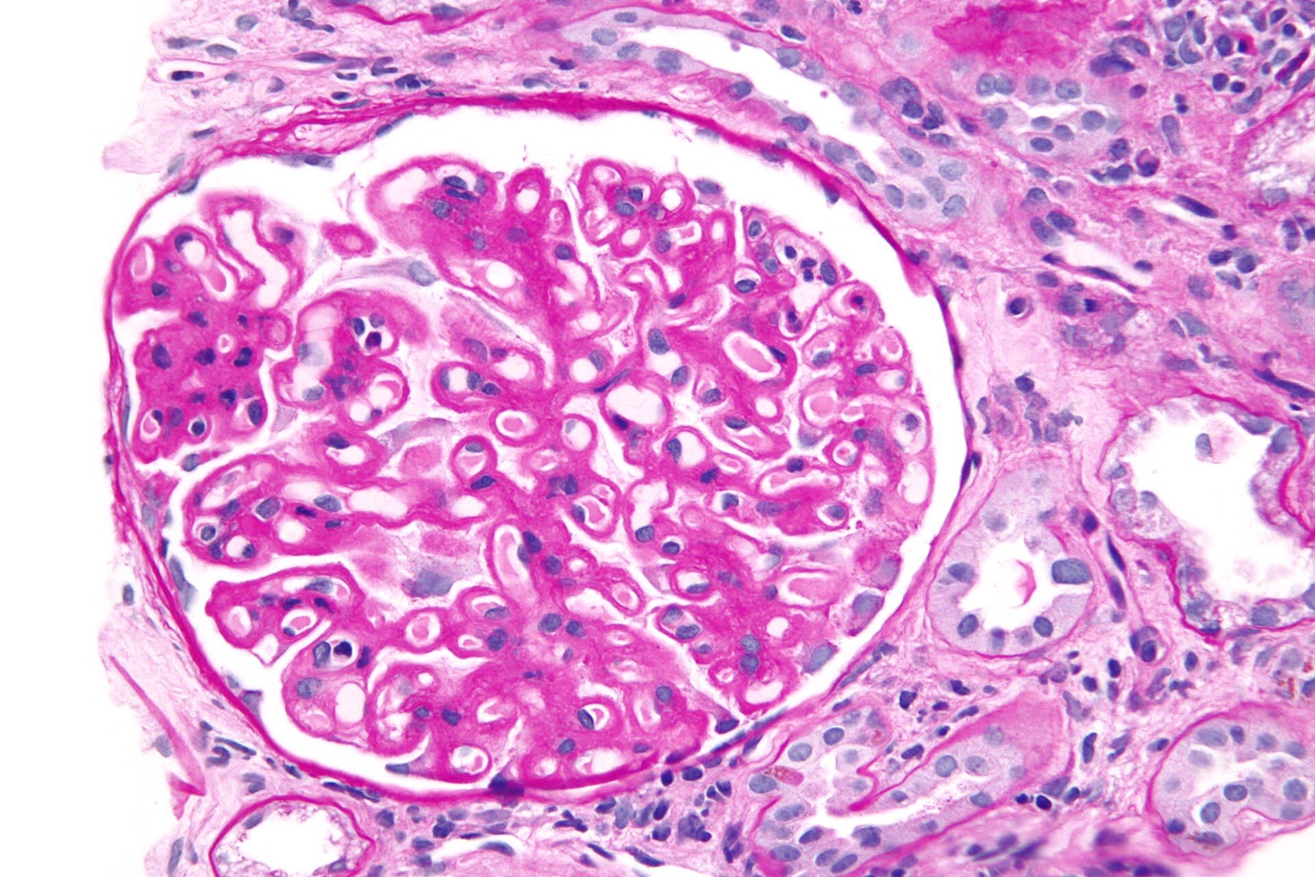
A renal biopsy from a patient with nephrotic syndrome demonstrates diffuse thickening of glomerular capillary walls on light microscopy. Which diagnosis is most consistent with this finding?
Nephron, CC BY-SA 3.0 <https://creativecommons.org/licenses/by-sa/3.0>, via Wikimedia Commons
Answer: B. Membranous nephropathy is characterized by diffuse thickening of glomerular capillary walls due to subepithelial immune complex deposition. Patients commonly present with nephrotic syndrome. Minimal change disease appears largely normal on light microscopy. FSGS produces focal sclerosis rather than diffuse thickening. IgA nephropathy is associated with mesangial proliferation and hematuria.
Source: Couser, W. G. (2017). Primary membranous nephropathy. Clinical Journal of the American Society of Nephrology.
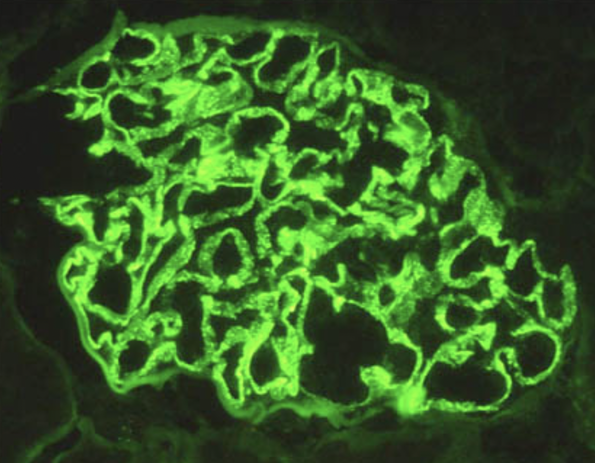
A renal biopsy demonstrates linear immunofluorescence staining along the glomerular basement membrane. Which disease is most consistent with this finding?
Adel.stoyanova, CC BY-SA 4.0 <https://creativecommons.org/licenses/by-sa/4.0>, via Wikimedia Commons
Answer: C. Goodpasture syndrome is characterized by antibodies directed against type IV collagen in basement membranes, producing linear immunofluorescence staining. IgA nephropathy shows mesangial IgA deposition, while poststreptococcal glomerulonephritis produces granular immune complex deposition. Minimal change disease lacks immune deposits on light microscopy. The linear staining pattern is highly specific for anti-GBM disease.
Source: McAdoo, et al. (2017). Pathogenesis and treatment of anti-GBM disease. Clinical Journal of the American Society of Nephrology.
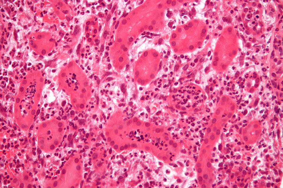
A 38-year-old woman presents with fever, flank pain, and costovertebral angle tenderness. A biopsy is shown below. What best explains this pattern?
Nephron, CC BY-SA 3.0 <https://creativecommons.org/licenses/by-sa/3.0>, via Wikimedia Commons
Answer: A. Acute pyelonephritis results from bacterial ascent from the bladder through the ureters into the renal pelvis and tubules. Bacterial products and cytokines recruit neutrophils into the tubules and interstitium, producing tubulitis and acute tubular injury. Glomeruli, not directly exposed to the ascending bacteria, are typically spared. Option B describes atypical hematogenous spread with primary glomerular injury. Options C–E misattribute the findings to immune-complex disease, chronic inflammation, or artifact rather than true acute bacterial pyelonephritis.
Source: Czaja, C. A., et al. (2007). Population-based epidemiologic analysis of acute pyelonephritis. Clinical Infectious Diseases, 45(3).
Your next questions are ready.
The question bank picks up from here: it adapts to what you just missed and brings it back right before you'd forget it.
What is the difference between PSGN and IgA nephropathy?expand_more
PSGN appears 1–3 weeks after strep pharyngitis (3–6 weeks after skin infection) with low C3. IgA nephropathy causes hematuria within days of an infection with normal complement and mesangial IgA deposits.
What are crescents?expand_more
Proliferations of parietal epithelial cells, macrophages, and fibrin in Bowman space. They define rapidly progressive glomerulonephritis.
Which glomerular diseases have low complement?expand_more
Poststreptococcal GN, lupus nephritis, membranoproliferative GN, and cryoglobulinemic GN.
What does Goodpasture syndrome target?expand_more
The α3 chain of type IV collagen in glomerular and alveolar basement membranes, producing linear IgG deposits.
Study smarter with a short weekly email. No spam — unsubscribe anytime.
✓ You're in — talk soon.
Reading is one thing. Remembering is another.
Turn this guide into questions you’ll actually remember
Progress Note turns concepts like this into adaptive questions and spaced review, so difficult topics come back to you exactly when you’re about to forget them.