Inflammation is the body's response to injury and infection: bring fluid and leukocytes to the site, destroy the offending agent, and clear the debris. Acute inflammation is fast and neutrophil-driven; chronic inflammation is slow, macrophage- and lymphocyte-driven, and mixes destruction with repair. Step 1 tests the steps of leukocyte recruitment, which mediator causes which sign, the cell that dominates each phase, and granulomas.
Evidence-based · For exam prep, not medical advice
update Updated October 2026
Date of last content change
Built by surgery and anesthesiology residents
Written for USMLE Step 1 · Free to read
bolt The 60-second version
Inflammation is the body's response to injury and infection: bring fluid and leukocytes to the site, destroy the offending agent, and clear the debris. Acute inflammation is fast and neutrophil-driven; chronic inflammation is slow, macrophage- and lymphocyte-driven, and mixes destruction with repair.
check_circleAcute inflammation: vasodilation and increased vascular permeability (edema), then neutrophil recruitment, within minutes to days.
check_circleLeukocyte recruitment: margination and rolling (selectins) → tight adhesion (integrins) → transmigration (PECAM-1) → chemotaxis.
check_circleHistamine causes immediate vasodilation and permeability; prostaglandins cause pain and fever; leukotriene B4 and IL-8 attract neutrophils.
check_circleNeutrophils dominate the first 24–48 hours; macrophages follow and drive resolution or chronic inflammation.
check_circleChronic inflammation features macrophages, lymphocytes, and plasma cells with simultaneous tissue destruction and fibrosis.
check_circleGranulomas are collections of epithelioid macrophages; caseating granulomas suggest tuberculosis or fungi, noncaseating suggest sarcoidosis or Crohn disease.
target What you'll be able to do · aligned to USMLE Step 1 · General Principles — Pathology & Immunology
PATHDescribe the vascular changes of acute inflammation and distinguish an exudate from a transudate.
PATHOutline the steps of leukocyte recruitment and the adhesion molecules that mediate each step.
PATHMatch the major chemical mediators of inflammation to their sources and effects.
IMMUNOContrast acute, chronic, and granulomatous inflammation and recognize neutrophil defects such as LAD and CGD.
How inflammation is tested on Step 1
Vignettes pair a clinical sign with a mediator, a defect in leukocyte recruitment with an immunodeficiency, or a biopsy with the type of inflammation. Leukocyte adhesion deficiency and chronic granulomatous disease are favorites.
workspace_premium The cardinal signs
Rubor (redness) and calor (heat) from vasodilation, tumor (swelling) from leaky vessels, dolor (pain) from prostaglandins and bradykinin, and functio laesa (loss of function).
Vascular changes
Within seconds of injury, arterioles briefly constrict, then dilate. Increased flow causes redness and warmth. Endothelial cells contract and open gaps in postcapillary venules, letting protein-rich fluid (exudate) escape into the tissue. Fluid loss concentrates red cells and slows flow (stasis), positioning leukocytes along the vessel wall.
Fluid
Protein and cells
Specific gravity
Cause
Exudate
High protein, many cells
> 1.020
Inflammation increases vascular permeability
Transudate
Low protein, few cells
< 1.012
Altered hydrostatic or oncotic pressure (heart failure, nephrotic syndrome)
An exudate signals inflammation; a transudate signals a pressure imbalance with intact vessels.
Leukocyte recruitment
Neutrophils leave the blood in a stepwise sequence, each step mediated by a specific family of adhesion molecules.
Leukocyte recruitment. Cytokines (TNF, IL-1) up-regulate selectins and integrin ligands on the endothelium; chemokines activate integrins on the leukocyte.
error Leukocyte adhesion deficiency
LAD type 1 is a defect in the CD18 integrin subunit — neutrophils cannot adhere tightly or leave the blood. Expect recurrent bacterial infections without pus, marked neutrophilia, and delayed separation of the umbilical cord.
Chemical mediators
Mediators come from cells (preformed or newly made) and from plasma proteins activated in cascades. Matching the mediator to its effect is high-yield.
Mediator
Source
Main effects
Histamine
Mast cells, basophils (preformed)
Immediate vasodilation and ↑ venular permeability
Prostaglandins (PGE2, PGI2)
Arachidonic acid via COX
Vasodilation, pain, fever (PGE2)
Leukotriene B4
Arachidonic acid via 5-lipoxygenase
Neutrophil chemotaxis and adhesion
Leukotrienes C4, D4, E4
5-lipoxygenase
Bronchospasm, vasoconstriction, ↑ permeability
Bradykinin
Kinin system (plasma)
↑ permeability, vasodilation, pain
C3a, C5a
Complement
Anaphylatoxins (mast-cell degranulation); C5a is a chemoattractant
NSAIDs block COX (prostaglandins) — reducing pain and fever but not leukotriene-mediated effects.
Time course: neutrophils, then macrophages
The cell population at the site changes predictably. Neutrophils arrive first because they are abundant and respond quickly; macrophages take over as the neutrophils die by apoptosis.
Cellular time course. Neutrophils predominate in the first 1–2 days; macrophages take over by days 2–3 and either resolve the response or sustain chronic inflammation.
workspace_premium Killing microbes
Neutrophils use the respiratory burst: NADPH oxidase makes superoxide, and myeloperoxidase converts H₂O₂ to bleach (HOCl). In chronic granulomatous disease, NADPH oxidase is defective — catalase-positive organisms (S. aureus, Aspergillus, Serratia, Nocardia) cause recurrent infections. The nitroblue tetrazolium test is negative.
Outcomes, chronic and granulomatous inflammation
Acute inflammation can resolve completely, heal by fibrosis, form an abscess, or progress to chronic inflammation when the agent persists.
Acute vs chronic inflammation. Chronic inflammation is defined by its cells and by simultaneous destruction and repair, not by duration alone.
Granulomas form when macrophages present antigen to CD4⁺ Th1 cells; IFN-γ converts macrophages to epithelioid cells and multinucleated giant cells. TNF maintains them — anti-TNF drugs can reactivate latent TB.
functions Worked example — name the defect
A 2-year-old has recurrent skin abscesses and pneumonia caused by Staphylococcus aureus and Aspergillus. His neutrophils migrate and phagocytose normally but fail to reduce nitroblue tetrazolium dye. What is the defect?
Normal migration and phagocytosis rule out an adhesion defect.
Failure to reduce NBT means no superoxide is produced — the respiratory burst is absent.
Catalase-positive organisms break down their own H₂O₂, so the neutrophil has no substrate to borrow and cannot kill them.
Answer. Chronic granulomatous disease — a defect of NADPH oxidase. Diagnosis is by the dihydrorhodamine (flow cytometry) or NBT test; treatment includes antimicrobial prophylaxis and interferon-γ.
Straight from the Progress Note question bank, each with a cited explanation. No account needed.
123
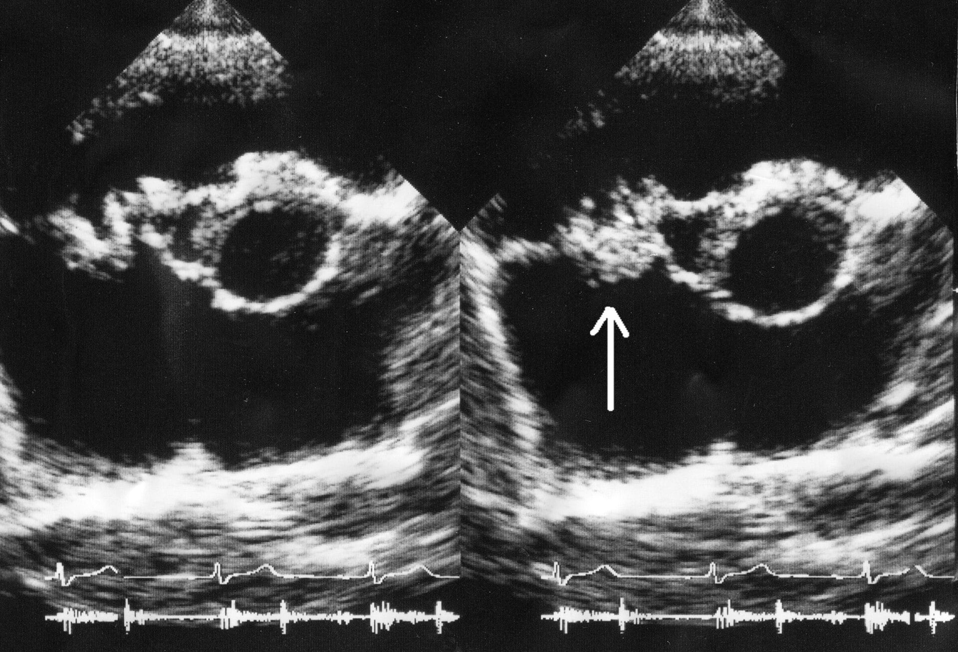
A 35-year-old man with a history of intravenous drug use presents with fever, night sweats, and a new holosystolic murmur loudest at the left lower sternal border that increases with inspiration. Blood cultures grow Staphylococcus aureus. Echocardiography reveals a vegetation on the tricuspid valve. Which of the following histological findings would be expected in the valve leaflet at the site of vegetation?
Endocarditis_ultrasound.gif: Daisuke Koya, Kazuyuki Shibuya, Ryuichi Kikkawa and Masakazu Haneda.derivative work: Materialscientist, CC BY 2.0 <https://creativecommons.org/licenses/by/2.0>, via Wikimedia Commons
Answer: B. Acute infective endocarditis caused by Staphylococcus aureus produces bulky, friable vegetations composed of fibrin, platelets, inflammatory cells, and dense bacterial colonies on damaged or previously normal valve surfaces. Histologically, the vegetation shows necrotic and ulcerated endothelium overlying thrombotic material with embedded gram-positive cocci, surrounded by acute neutrophilic infiltrate — reflecting the highly virulent, tissue-destructive nature of S. aureus. Right-sided endocarditis (tricuspid valve) in IV drug users is characteristic because skin flora enter directly into the venous circulation. Sterile platelet-fibrin thrombi without inflammation (option A) describe non-bacterial thrombotic (marantic) endocarditis, seen in hypercoagulable states and malignancy. Aschoff bodies (option D) are pathognomonic for rheumatic carditis, caused by cross-reactive immune injury to the mitral valve.
Source: Murdoch, et al. (2009). Clinical presentation, etiology, and outcome of infective endocarditis in the 21st century. Archives of Internal Medicine, 169(5), 463–473.
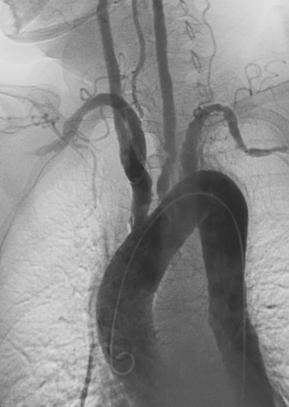
A 25-year-old woman of Asian descent has weak or absent radial pulses bilaterally, arm claudication, discordant blood pressures between limbs, and elevated inflammatory markers. Imaging of the aortic arch and its major branches is shown below. Which statement about the pathogenesis and typical demographic of this condition is correct?
Justin Ly, Public domain, via Wikimedia Commons
Answer: A. Takayasu arteritis is a cell-mediated (T-cell driven), granulomatous large-vessel vasculitis that predominantly affects the aorta and its major branches, typically presenting in young women, particularly of Asian descent, under the age of 40; the disease progresses from an early active inflammatory phase, with patchy granulomatous infiltration of the vessel wall, to a later fibrotic phase characterized by intimal and medial fibrosis producing luminal stenosis (explaining diminished pulses and limb claudication) and occasionally aneurysmal dilation. Option B incorrectly substitutes an immune complex-mediated mechanism more characteristic of polyarteritis nodosa, a distinct medium-vessel vasculitis. Option C incorrectly reverses both the typical demographic (older men rather than young women) and the underlying mechanism (atherosclerosis rather than autoimmune vasculitis). Option D incorrectly proposes an infectious mechanism rather than the autoimmune, cell-mediated process actually responsible. Option E incorrectly substitutes an IgA-mediated mechanism unrelated to this disease's pathogenesis.
Source: Mason, J. C. (2010). Takayasu arteritis—Advances in diagnosis and management. Nature Reviews Rheumatology, 6(7).
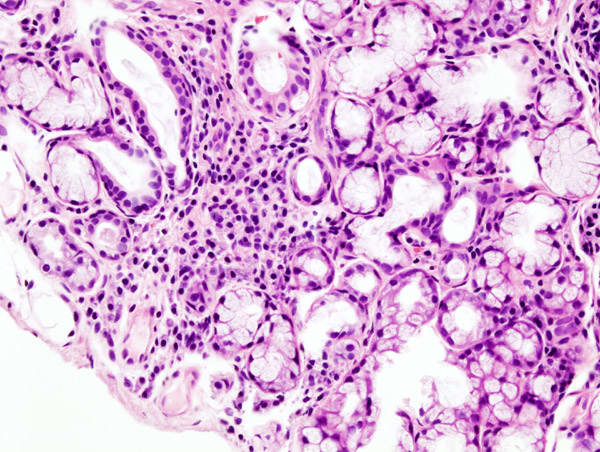
A 45-year-old woman has dry eyes, dry mouth, and recurrent dental caries. Serum anti-Ro/SSA and anti-La/SSB antibodies are positive. A minor salivary gland (lip) biopsy is performed, photo shown below. Which finding is consistent with the diagnosis, and what does it reflect about the underlying pathogenesis?
KGH, CC BY-SA 3.0 <http://creativecommons.org/licenses/by-sa/3.0/>, via Wikimedia Commons
Answer: B. Primary Sjögren's syndrome is characterized histologically by focal lymphocytic sialadenitis on minor salivary gland biopsy, in which dense periductal and perivascular aggregates of T and B lymphocytes progressively replace normal acinar architecture, reflecting an autoimmune, lymphocyte-mediated destruction of exocrine glandular tissue that underlies the clinical dryness (sicca symptoms); this finding, combined with anti-Ro/SSA and anti-La/SSB seropositivity, supports the diagnosis per current classification criteria. Option A incorrectly substitutes a granulomatous, caseating pattern characteristic of mycobacterial infection. Option C incorrectly attributes the findings to amyloid deposition from a plasma cell dyscrasia, an unrelated process. Option D incorrectly omits the lymphocytic infiltrate that is the defining histologic feature of this disease. Option E incorrectly proposes an acute bacterial process rather than the chronic autoimmune lymphocytic infiltrate actually observed.
Source: Mariette, X., & Criswell, L. A. (2018). Primary Sjögren's syndrome. New England Journal of Medicine.
Your next questions are ready.
The question bank picks up from here: it adapts to what you just missed and brings it back right before you'd forget it.
What are the steps of leukocyte extravasation?expand_more
Margination and rolling mediated by selectins, tight adhesion mediated by integrins binding ICAM-1, transmigration through endothelial junctions mediated by PECAM-1, and chemotaxis toward signals such as C5a, LTB4, and IL-8.
Which cell predominates in acute versus chronic inflammation?expand_more
Neutrophils predominate in acute inflammation, peaking at 24–48 hours. Chronic inflammation is dominated by macrophages, lymphocytes, and plasma cells.
What is the difference between caseating and noncaseating granulomas?expand_more
Caseating granulomas have central necrosis and suggest tuberculosis or systemic fungal infection. Noncaseating granulomas lack necrosis and suggest sarcoidosis, Crohn disease, berylliosis, or a foreign body.
Which mediators cause fever and pain?expand_more
Prostaglandin E2 causes fever (acting on the hypothalamus) and, with bradykinin, pain. IL-1 and TNF drive fever by stimulating prostaglandin production.
Study smarter with a short weekly email. No spam — unsubscribe anytime.
✓ You're in — talk soon.
Reading is one thing. Remembering is another.
Turn this guide into questions you’ll actually remember
Progress Note turns concepts like this into adaptive questions and spaced review, so difficult topics come back to you exactly when you’re about to forget them.