Cell injury is what happens when a stress exceeds a cell's ability to adapt. Early injury is reversible — the cell swells but can recover. Past a "point of no return," membranes fail, mitochondria collapse, and the cell dies by necrosis. Step 1 tests the mechanisms (ATP depletion, calcium, free radicals), the morphologic line between reversible and irreversible injury, and which pattern of necrosis appears in which organ. This guide walks the sequence from insult to cell death.
Evidence-based · For exam prep, not medical advice
update Updated October 2026
Date of last content change
Built by surgery and anesthesiology residents
Written for USMLE Step 1 · Free to read
bolt The 60-second version
Cell injury is what happens when a stress exceeds a cell's ability to adapt. Early injury is reversible — the cell swells but can recover. Past a "point of no return," membranes fail, mitochondria collapse, and the cell dies by necrosis.
check_circleHypoxia is the most common cause of cell injury; its central consequence is ATP depletion.
check_circleIrreversible injury = plasma membrane damage (enzyme leakage), lysosomal rupture, mitochondrial permeability with cytochrome c release, and nuclear destruction.
check_circleNuclear changes of cell death progress from pyknosis (shrinkage) to karyorrhexis (fragmentation) to karyolysis (fading).
check_circleCoagulative necrosis is the default for ischemia of solid organs; the brain is the exception — it undergoes liquefactive necrosis.
check_circleDystrophic calcification occurs in damaged tissue with normal serum calcium; metastatic calcification occurs in normal tissue when calcium or phosphate is high.
target What you'll be able to do · aligned to USMLE Step 1 · General Principles — Pathology
PATHDescribe how hypoxia, ATP depletion, calcium influx, and free radicals injure cells.
PATHDistinguish the morphologic features of reversible and irreversible cell injury, including the sequence of nuclear changes.
PATHMatch each pattern of necrosis to its mechanism and classic tissue or disease.
PATHDifferentiate dystrophic from metastatic calcification by serum calcium and tissue setting.
How cell injury is tested on Step 1
Questions usually hand you a tissue and a mechanism — an ischemic heart, a brain infarct, a tuberculous lung, a necrotic pancreas — and ask you to name the morphologic pattern, the reversible-versus-irreversible change shown, or the enzyme that will rise in the blood.
workspace_premium The through-line
Almost every cause of injury converges on a few final pathways: ATP depletion, mitochondrial damage, calcium influx, free radicals, and membrane damage. Membrane damage plus mitochondrial failure marks the point of no return.
Mechanisms: the final common pathways
Causes of injury include hypoxia and ischemia (most common), toxins and drugs, infections, immune reactions, physical agents, genetic defects, and nutritional imbalance. Whatever the cause, damage flows through a small set of shared mechanisms.
ATP depletion drives early injury. Without ATP, the Na⁺/K⁺-ATPase fails, sodium and water flood in, and the cell and its organelles swell. A parallel shift to anaerobic glycolysis builds lactic acid, lowers pH, and clumps nuclear chromatin.
Mechanism
What goes wrong
Classic example
ATP depletion
Ion pumps fail, protein synthesis stops, anaerobic glycolysis lowers pH
Myocardial ischemia
Mitochondrial damage
Loss of membrane potential; cytochrome c leaks and can trigger apoptosis
Hypoxia, toxins
Calcium influx
Activates phospholipases, proteases, endonucleases, and ATPases
Ischemia-reperfusion
Free radicals
Lipid peroxidation, protein oxidation, DNA breaks
Reperfusion, radiation, acetaminophen
Membrane damage
Loss of selective permeability; enzymes leak out
Irreversible injury of any cause
Each pathway feeds the others — ATP loss allows calcium in, calcium damages mitochondria, and damaged mitochondria generate free radicals.
Reversible vs irreversible injury
The difference is whether the cell can recover once the stimulus is removed. Reversible changes reflect a cell that is struggling; irreversible changes reflect structures that cannot be repaired.
The point of no return. Two findings define irreversibility: inability to restore mitochondrial function, and severe disturbance of membrane function.Nuclear changes of cell death occur in this order and are the most reliable light-microscopic sign that a cell is dead.
error Why serum enzymes rise
Membrane damage lets cytoplasmic proteins leak into the blood — the basis of clinical injury markers: troponin (myocardium), ALT/AST (liver), lipase (pancreas), and CK (muscle).
Free radical injury and reperfusion
Free radicals are unstable molecules with an unpaired electron that damage lipids, proteins, and DNA. They are generated by normal metabolism and amplified in disease; the cell neutralizes them with enzymes and antioxidant vitamins.
Sources of free radicals
Defenses
Ionizing radiation (hydrolysis of water → hydroxyl radical)
Superoxide dismutase
Inflammation: NADPH oxidase in neutrophils (respiratory burst)
Injury occurs when generation outpaces these defenses.
workspace_premium Reperfusion injury
Restoring blood flow to ischemic tissue can paradoxically worsen damage: returning oxygen generates a burst of free radicals, calcium floods damaged cells, and neutrophils arrive and amplify inflammation. This is why troponin can rise further right after a successful reperfusion.
The six patterns of necrosis
Necrosis is unregulated cell death that ruptures membranes and provokes inflammation. Its appearance depends on whether protein denaturation or enzymatic digestion dominates — which in turn depends on the tissue and the cause.
Pattern
Mechanism and appearance
Classic setting
Coagulative
Proteins denature; cell outlines and tissue architecture preserved ("ghost cells")
Ischemic infarcts of the heart, kidney, spleen
Liquefactive
Enzymatic digestion turns tissue into a liquid mass
Brain infarcts; bacterial abscesses
Caseous
Cheese-like, friable debris inside a granuloma
Tuberculosis; systemic fungi
Fat
Lipases free fatty acids, which bind calcium (saponification)
Acute pancreatitis (enzymatic); breast trauma
Fibrinoid
Immune complexes and fibrin deposit in vessel walls
Vasculitis, malignant hypertension, preeclampsia
Gangrenous
Coagulative necrosis of a limb; "wet" if infection adds liquefaction
Diabetic foot, limb ischemia
The brain is the key exception to the ischemia rule: its high lipid content and lysosomal enzymes produce liquefactive, not coagulative, necrosis.
error Common trap
An ischemic infarct in the brain is liquefactive; an ischemic infarct in the heart is coagulative. Both are ischemic — the tissue decides the pattern.
Pathologic calcification
Calcium salts deposit in tissues in two settings that Step 1 distinguishes by one question: is the serum calcium normal?
Dystrophic vs metastatic calcification. Metastatic calcification favors tissues that lose acid and become relatively alkaline — the kidney, lung, and stomach.
functions Worked example — name the process
A 48-year-old man with heavy alcohol use dies of acute pancreatitis. At autopsy, the peripancreatic fat contains chalky white deposits. Serum calcium was low on admission. What process produced the deposits?
Pancreatic lipases released into the surrounding fat break triglycerides into free fatty acids.
The free fatty acids bind calcium, forming chalky white soaps (saponification).
The deposits form in damaged tissue while serum calcium is normal or low — so this is dystrophic, not metastatic, calcification.
Answer. Enzymatic fat necrosis with saponification — a form of dystrophic calcification. Consumption of calcium in saponification also explains the hypocalcemia seen in severe pancreatitis.
Straight from the Progress Note question bank, each with a cited explanation. No account needed.
12
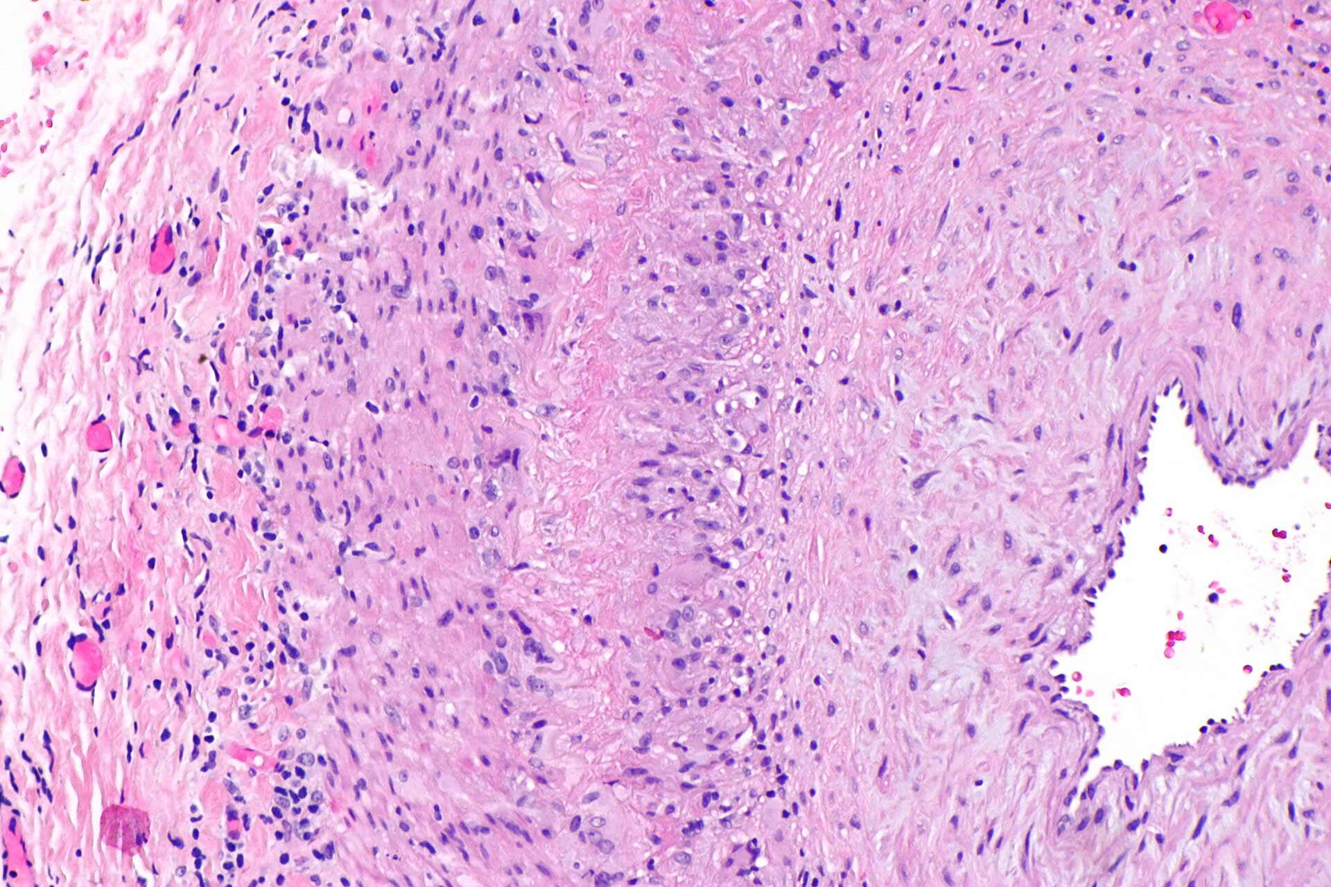
A 72-year-old woman presents with new-onset temporal headache, jaw claudication while chewing, scalp tenderness, and blurred vision in one eye. ESR is markedly elevated at 95 mm/hr. Temporal artery biopsy, photomicrograph shown below, reveals granulomatous inflammation of the vessel wall with multinucleated giant cells and fragmentation of the internal elastic lamina. Which of the following best describes the mechanism responsible for this patient's arterial wall damage and risk of irreversible vision loss?
Nephron, CC BY-SA 3.0 <https://creativecommons.org/licenses/by-sa/3.0>, via Wikimedia Commons
Answer: A. Giant cell arteritis is a granulomatous vasculitis of medium and large arteries, particularly cranial branches of the carotid artery, mediated by CD4+ T cells and macrophages that infiltrate the vessel wall and form multinucleated giant cells; this inflammatory process destroys the internal elastic lamina and can compromise flow through the posterior ciliary arteries supplying the optic nerve, producing irreversible anterior ischemic optic neuropathy if untreated. This is why high-dose glucocorticoids are started immediately upon clinical suspicion, without waiting for biopsy confirmation. ANCA-associated immune complex deposition describes a distinct category of small-vessel vasculitis, and atherosclerotic plaque rupture is a degenerative rather than granulomatous inflammatory process, making both mechanistically unrelated to this patient's disease.
Source: Weyand, C. M., & Goronzy, J. J. (2014). Giant-cell arteritis and polymyalgia rheumatica. New England Journal of Medicine, 371(1).
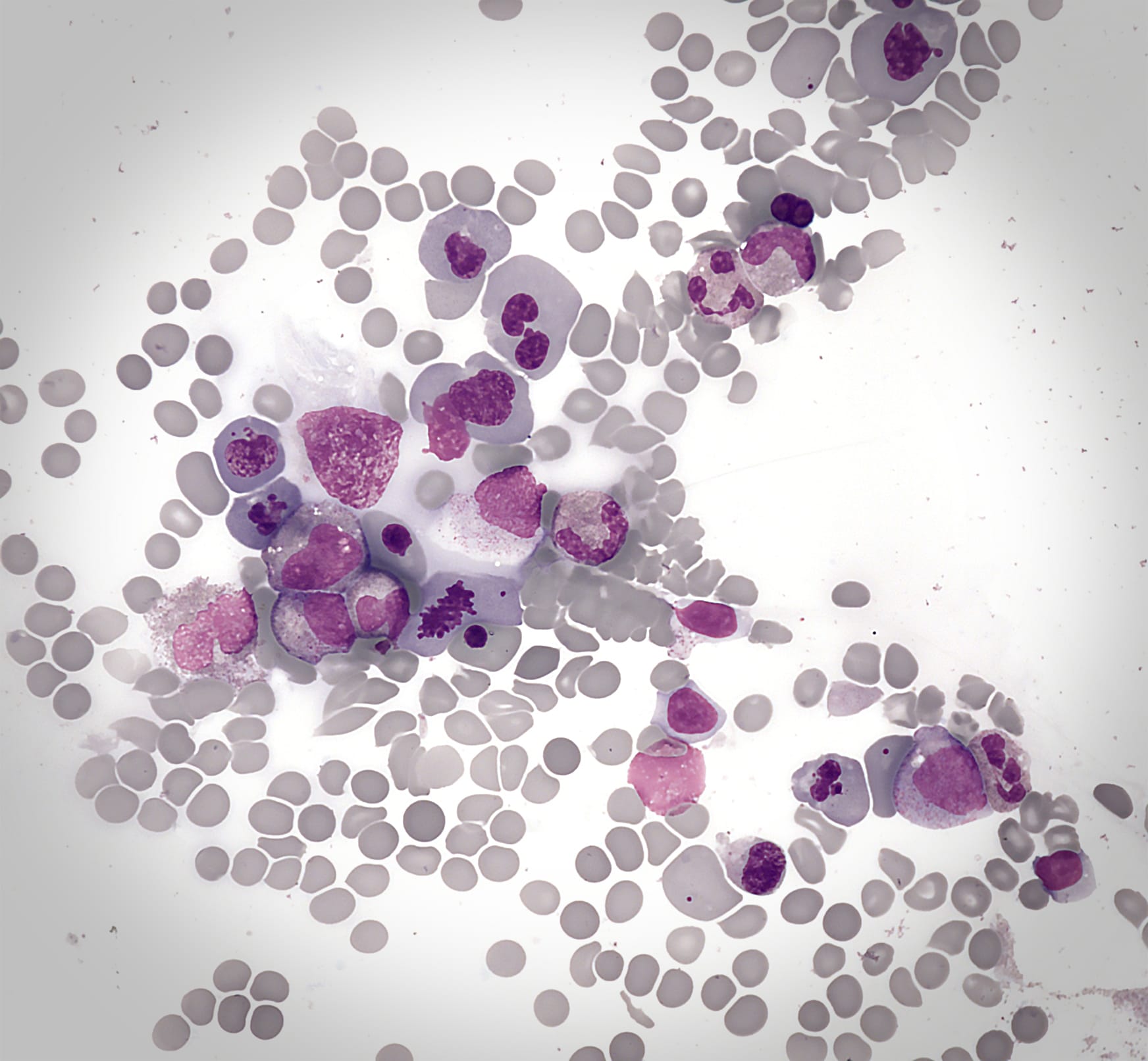
A patient receiving chronic methotrexate therapy for rheumatoid arthritis develops oral ulcers and pancytopenia. Bone marrow biopsy demonstrates megaloblastic changes with enlarged erythroid precursor cells. Which mechanism best explains these findings?
Makysm, CC0, via Wikimedia Commons
Answer: A. Methotrexate inhibits dihydrofolate reductase, reducing tetrahydrofolate production and impairing thymidine synthesis. Rapidly dividing cells in the bone marrow are particularly affected, resulting in megaloblastic changes and pancytopenia. Inosine monophosphate dehydrogenase is inhibited by mycophenolate, while topoisomerase II is targeted by etoposide. The histologic findings reflect impaired DNA synthesis due to folate antagonism.
Source: Cronstein, et al. (2020). Methotrexate and its mechanisms of action in inflammatory arthritis. Nature Reviews Rheumatology.
Your next questions are ready.
The question bank picks up from here: it adapts to what you just missed and brings it back right before you'd forget it.
What is the difference between reversible and irreversible cell injury?expand_more
Reversible injury shows cellular swelling, fatty change, membrane blebbing, and chromatin clumping, and the cell recovers if the stimulus is removed. Irreversible injury shows plasma membrane damage with enzyme leakage, lysosomal rupture, mitochondrial failure, and nuclear destruction (pyknosis, karyorrhexis, karyolysis).
Why does the brain undergo liquefactive necrosis instead of coagulative necrosis?expand_more
Brain tissue is rich in lipids and lysosomal enzymes and has little supporting stroma, so enzymatic digestion dominates over protein denaturation after ischemia, turning the infarct into a liquefied cavity.
What is the most common cause of cell injury?expand_more
Hypoxia, most often from ischemia. Its central effect is depletion of ATP, which causes failure of the sodium-potassium pump and cell swelling.
What causes reperfusion injury?expand_more
When blood flow returns, oxygen generates a burst of free radicals, calcium enters damaged cells, and neutrophils amplify inflammation, extending damage beyond the original ischemic injury.
Study smarter with a short weekly email. No spam — unsubscribe anytime.
✓ You're in — talk soon.
Reading is one thing. Remembering is another.
Turn this guide into questions you’ll actually remember
Progress Note turns concepts like this into adaptive questions and spaced review, so difficult topics come back to you exactly when you’re about to forget them.