Apoptosis is programmed, energy-dependent cell death. Unlike necrosis, it is orderly: the cell shrinks, its chromatin condenses, it breaks into membrane-bound fragments, and macrophages clear it without inflammation. Step 1 tests the two triggering pathways, the proteins that control them, the morphology, and the diseases that result when apoptosis is too strong or too weak.
Evidence-based · For exam prep, not medical advice
update Updated October 2026
Date of last content change
Built by surgery and anesthesiology residents
Written for USMLE Step 1 · Free to read
bolt The 60-second version
check_circleApoptosis is ATP-dependent, programmed cell death that does not cause inflammation.
check_circleThe intrinsic (mitochondrial) pathway is controlled by the BCL-2 family: BCL-2 and BCL-xL block it; BAX and BAK open the mitochondrial membrane to release cytochrome c.
check_circleThe extrinsic pathway is triggered by death receptors — Fas ligand binding Fas (CD95) or TNF binding its receptor — and activates caspase-8.
check_circleBoth pathways converge on executioner caspases (caspase-3), which cleave proteins and activate endonucleases.
check_circleMorphology: cell shrinkage, pyknosis, karyorrhexis, membrane blebbing, and apoptotic bodies cleared by macrophages; DNA laddering on electrophoresis.
check_circleToo little apoptosis drives cancer (BCL-2 overexpression in follicular lymphoma) and autoimmunity; too much contributes to neurodegeneration and ischemic injury.
target What you'll be able to do · aligned to USMLE Step 1 · General Principles — Pathology
PATHDescribe the intrinsic and extrinsic apoptotic pathways and the caspases that execute them.
PATHExplain how the BCL-2 family and p53 regulate mitochondrial apoptosis.
PATHRecognize the morphologic and laboratory features of apoptosis and contrast them with necrosis.
PATHRelate defects of apoptosis to cancer, autoimmunity, and neurodegeneration.
How apoptosis is tested on Step 1
Questions ask which protein is mutated or overexpressed, which pathway a cytotoxic T cell uses, which finding distinguishes apoptosis from necrosis, or which disease results from failed apoptosis.
workspace_premium The through-line
Two doors lead in — the mitochondria (intrinsic, sensing internal damage) and the death receptors (extrinsic, signals from other cells) — and both open onto the same caspase cascade.
The intrinsic (mitochondrial) pathway
The intrinsic pathway responds to internal stress: DNA damage, loss of growth-factor or hormonal signals, misfolded proteins, hypoxia, and radiation. Its gatekeeper is the BCL-2 family, which controls the permeability of the outer mitochondrial membrane.
Intrinsic pathway. Pro-apoptotic BAX and BAK form channels in the outer mitochondrial membrane; cytochrome c then binds APAF-1 to form the apoptosome, which activates caspase-9.
Protein
Role
Clinical link
BCL-2, BCL-xL
Anti-apoptotic — keep the mitochondrial membrane closed
Overexpressed in follicular lymphoma t(14;18)
BAX, BAK
Pro-apoptotic effectors — create channels for cytochrome c
Activated when stress outweighs BCL-2
BH3-only (BIM, BID, PUMA)
Sensors of stress; neutralize BCL-2
BID links the extrinsic pathway to mitochondria
p53
Senses DNA damage; induces PUMA and BAX
Mutated in about half of cancers (Li-Fraumeni when germline)
The balance between pro- and anti-apoptotic BCL-2 family members decides whether the cell lives.
The extrinsic (death receptor) pathway
The extrinsic pathway is triggered from outside the cell when a ligand binds a death receptor of the TNF-receptor family. It is how the immune system removes unwanted lymphocytes and infected or tumor cells.
Extrinsic pathway. Death-receptor engagement recruits FADD and activates caspase-8. Caspase-8 can also cleave BID, recruiting the mitochondrial pathway to amplify the signal.
Fas–FasL: deletes self-reactive lymphocytes; defects cause autoimmune lymphoproliferative syndrome (ALPS).
Cytotoxic T cells kill targets by FasL and by releasing perforin and granzyme B, which enters the cell and activates caspases directly.
Executioner caspases cleave the cytoskeleton and nuclear lamins and activate endonucleases that cut DNA between nucleosomes. The cell packages itself for clean removal.
Cell shrinkage with dense, eosinophilic cytoplasm.
Chromatin condensation (pyknosis) — the most characteristic feature — then nuclear fragmentation (karyorrhexis).
Membrane blebbing and formation of apoptotic bodies — cytoplasm and nuclear fragments wrapped in intact membrane.
Phagocytosis: phosphatidylserine flips to the outer leaflet, signaling macrophages to engulf the bodies without inflammation.
workspace_premium Lab correlate
Internucleosomal cleavage produces DNA fragments in multiples of ~180 base pairs — the "DNA ladder" on gel electrophoresis. Necrosis produces a diffuse smear instead.
Apoptosis vs necrosis
The two forms of cell death differ in control, energy use, and whether the neighbors notice.
Apoptosis vs necrosis. Membrane integrity is the clearest dividing line.
Setting
Example
Embryogenesis
Removal of webbing between digits; involution of the Müllerian duct by AMH in males
Hormone withdrawal
Endometrial shedding during menstruation
Immune regulation
Negative selection of self-reactive T cells in the thymus
Cell turnover
Crypt cells of the intestine
Pathologic triggers
Viral hepatitis (Councilman bodies), DNA damage from radiation or chemotherapy
Apoptosis is part of normal development as well as disease.
functions Worked example — follow the protein
A 50-year-old has painless lymphadenopathy. Biopsy shows a follicular lymphoma with a t(14;18) translocation. How does this translocation promote tumor growth?
t(14;18) places the BCL2 gene next to the immunoglobulin heavy-chain promoter on chromosome 14.
B cells therefore overexpress BCL-2, an anti-apoptotic protein.
BCL-2 keeps the mitochondrial membrane closed, so damaged B cells fail to undergo intrinsic apoptosis and accumulate.
Answer. Overexpression of anti-apoptotic BCL-2 blocks the intrinsic pathway, so lymphoma cells survive rather than proliferate unusually fast — which is why follicular lymphoma is typically indolent.
Straight from the Progress Note question bank, each with a cited explanation. No account needed.
123
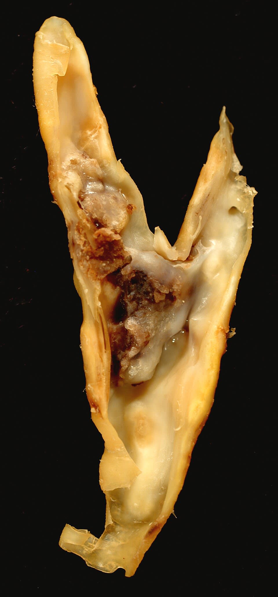
A 67-year-old man with a history of hypertension and hyperlipidemia undergoes carotid endarterectomy. The resected specimen is sent to pathology. Gross examination reveals a raised, yellow-white plaque with a necrotic core, shown in the image below. Which of the following cell types is most responsible for the foam cell formation the pathologist would see in this lesion?
Ed Uthman, CC BY 2.0 <https://creativecommons.org/licenses/by/2.0>, via Wikimedia Commons
Answer: D. Foam cells are the histologic hallmark of early and advanced atherosclerotic plaques and are derived predominantly from macrophages that have migrated into the intima and engulfed oxidized LDL (oxLDL) through scavenger receptors (SR-A and CD36), which—unlike the LDL receptor—are not subject to downregulation by intracellular cholesterol accumulation. This unregulated uptake leads to massive intracellular lipid accumulation, giving the cells their characteristic foamy cytoplasmic appearance on H&E. Smooth muscle cells (option A) can also become foam cells but are not the primary contributor. Option C is incorrect; neutrophils do not primarily contribute to foam cell formation and do not express scavenger receptors in this context. Option B is incorrect; T lymphocytes do not internalize lipoproteins to form foam cells. Option E is not a recognized mechanism of foam cell formation.
Source: Moore, K. J., & Tabas, I. (2011). Macrophages in the pathogenesis of atherosclerosis. Cell, 145(3), 341–355.
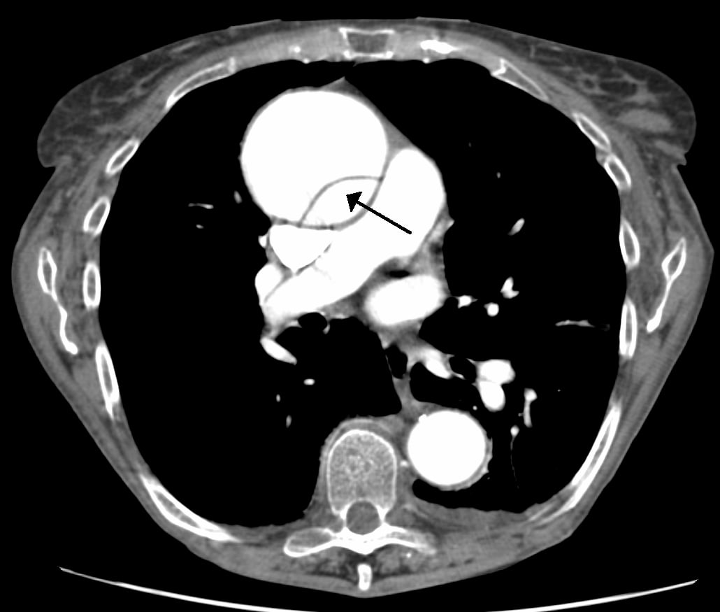
A 58-year-old man with connective tissue disease (Marfan syndrome) presents with acute tearing chest pain radiating to the back. CT angiography of the chest is shown below. Histology of the aortic wall shows fragmentation and disorganization of elastic fibers in the tunica media with areas of apparent elastic fiber loss (cystic medial degeneration). Which of the following best explains the pathophysiologic relationship between fibrillin-1 deficiency in Marfan syndrome, structural weakness of the aortic media, and the pathology observed in this patient?
James Heilman, MD, CC BY-SA 3.0 <https://creativecommons.org/licenses/by-sa/3.0>, via Wikimedia Commons
Answer: B. In Marfan syndrome, fibrillin-1 mutations impair the formation of extracellular microfibrils that normally sequester and stabilize latent TGF-beta complexes. Loss of sequestration results in excessive bioavailable TGF-beta signaling on aortic smooth muscle cells via TGF-beta receptors; chronic excessive TGF-beta signaling promotes smooth muscle cell apoptosis, impairs elastic fiber cross-linking, and inhibits smooth muscle proliferation recovery. The result is cystic medial degeneration—progressive loss and fragmentation of elastic fibers with smooth muscle cell death, creating a structurally weakened aortic media. This degeneration is reflected histologically as focal elastic fiber loss, disorganization, and cystic spaces. The weakened aortic media is predisposed to dissection when acute hemodynamic stress (hypertension, increased contractility, shear stress) exceeds the residual tensile strength of the media. The dissection initiates when an intimal tear allows blood to track into the media, separating the intima from the adventitia along a plane of medial weakness. Option A oversimplifies the mechanism; option C misattributes deficiency to collagen rather than elastic fibers; option D inverts the TGF-beta effect; option E incorrectly attributes degeneration to proteoglycan loss.
Source: Isselbacher, E. M., et al. (2022). 2022 ACC/AHA guideline for the diagnosis and management of aortic disease. Journal of the American College of Cardiology, 80(24).
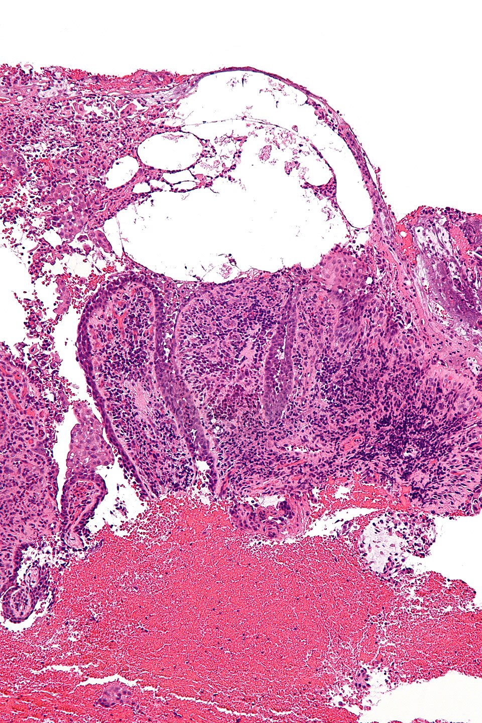
A 34-year-old woman presents with a 2-week history of recurrent painful vesicles and erosions on the lips, gums, tongue, and buccal mucosa, with a positive Nikolsky sign on intact oral mucosa. Intraoral biopsy is shown below. Which of the following best explains the pathophysiology and how autoimmune dysfunction of desmoglein-3 leads to the characteristic blistering observed in this patient?
Nephron, CC BY-SA 3.0 <https://creativecommons.org/licenses/by-sa/3.0>, via Wikimedia Commons
Answer: B. Pemphigus vulgaris is an autoimmune disorder in which IgG autoantibodies target desmoglein-3 (Dsg3), a transmembrane glycoprotein in desmosomes that mediates cell-to-cell adhesion. IgG antibodies bind Dsg3 and cause desmosomal dysfunction through multiple mechanisms: antibody binding induces conformational changes in desmoglein-3, disrupts calcium-dependent Dsg3-Dsg3 and Dsg3-desmocollin interactions, and triggers complement-mediated injury of the desmosomal complex. The disruption of desmosomal adhesion causes acantholysis—loss of intercellular cohesion and detachment of cells from one another. Histologically, this appears as intraepidermal blistering with preservation of the basal cell layer attached to the basement membrane (basal cells appear as a single layer at the blister base, creating the "tombstone" appearance). Direct immunofluorescence reveals IgG deposits distributed intercellularly in a "chicken-wire" pattern, reflecting the distribution of the desmoglein-3 antigen on the cell surface. The positive Nikolsky sign—exfoliation of superficial epidermis with light friction—reflects the widespread loss of cell-to-cell adhesion in intact epidermis from circulating IgG antibodies. Oral mucosa is frequently the initial site of involvement because Dsg3 is highly expressed in the oral epithelium. The severity of pemphigus correlates with serum anti-Dsg3 antibody titers. Option A incorrectly attributes the process to mast cell activation; option C misidentifies the target antigen (hemidesmosomes vs desmosomes); option D confuses acantholysis with apoptosis; option E reverses the primary and secondary processes.
Source: Schmidt, E., et al. (2019). Pemphigus. The Lancet, 394(10201).
Your next questions are ready.
The question bank picks up from here: it adapts to what you just missed and brings it back right before you'd forget it.
What is the difference between the intrinsic and extrinsic pathways of apoptosis?expand_more
The intrinsic pathway responds to internal stress such as DNA damage; BAX and BAK release cytochrome c from mitochondria, activating caspase-9. The extrinsic pathway starts when a ligand such as Fas ligand or TNF binds a death receptor, activating caspase-8. Both activate executioner caspase-3.
Why does apoptosis not cause inflammation?expand_more
The cell membrane stays intact, so contents do not spill into the tissue. Apoptotic bodies display phosphatidylserine and are engulfed by macrophages before they can trigger an inflammatory response.
What does BCL-2 do?expand_more
BCL-2 is an anti-apoptotic protein that keeps the outer mitochondrial membrane closed, preventing cytochrome c release. Overexpression, as in follicular lymphoma with t(14;18), lets damaged cells survive.
What is a DNA ladder?expand_more
In apoptosis, endonucleases cut DNA between nucleosomes into fragments in multiples of about 180 base pairs, which appear as a ladder on gel electrophoresis. Necrosis produces a diffuse smear.
Study smarter with a short weekly email. No spam — unsubscribe anytime.
✓ You're in — talk soon.
Reading is one thing. Remembering is another.
Turn this guide into questions you’ll actually remember
Progress Note turns concepts like this into adaptive questions and spaced review, so difficult topics come back to you exactly when you’re about to forget them.