How brain tumors are tested on Step 1
Age first, then location, then histology.
Adult tumors
Location and histology identify the tumor.
| Tumor | Location and imaging | Histology and markers |
|---|---|---|
| Metastases | Multiple, gray-white junction, edema | Resemble the primary cancer |
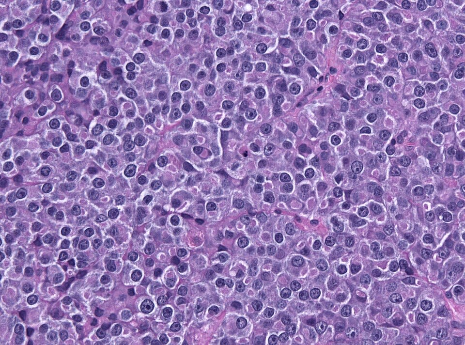
| Glioblastoma | Cerebral hemisphere; crosses corpus callosum; ring enhancing | Pseudopalisading necrosis, vascular proliferation; GFAP+ |
| Oligodendroglioma | Frontal lobe; calcified; seizures | "Fried egg" cells, "chicken wire" capillaries; 1p/19q codeletion |
| Meningioma | Dural-based, compresses brain; dural tail | Whorls, psammoma bodies |
| Vestibular schwannoma | Cerebellopontine angle | Antoni A and B areas, Verocay bodies; S-100+ |
| Hemangioblastoma | Cerebellum; von Hippel-Lindau | Foamy cells, capillaries; can make EPO (polycythemia) |
| Pituitary adenoma | Sella; bitemporal hemianopia | Prolactinoma most common |
Primary brain tumors rarely spread outside the CNS.
Pediatric tumors
Most arise in the posterior fossa and cause ataxia or hydrocephalus.
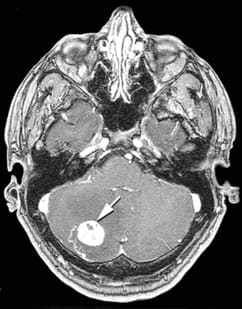
- Pilocytic astrocytoma: most common benign pediatric brain tumor; cerebellar cyst with a solid nodule; Rosenthal fibers; excellent prognosis after resection.
- Medulloblastoma: malignant tumor of the cerebellar vermis; truncal ataxia; small round blue cells with Homer Wright rosettes; can spread through CSF ("drop metastases").
- Ependymoma: fourth ventricle; blocks CSF flow (hydrocephalus); perivascular pseudorosettes.
- Craniopharyngioma: above the sella from Rathke pouch remnants; calcified cyst with "motor oil" fluid; bitemporal hemianopia, growth failure.
Raised intracranial pressure and herniation
A growing mass shifts brain tissue past rigid dural folds.
Worked example
Age, site, and histology.
functions Worked example — a child with clumsy walking
An 8-year-old has headaches and a wide-based, unsteady gait. MRI shows a cystic mass in the cerebellar hemisphere with an enhancing mural nodule. Biopsy shows eosinophilic, corkscrew-shaped fibers. What is the tumor and the prognosis?
- A child with a posterior fossa mass suggests pilocytic astrocytoma, medulloblastoma, or ependymoma.
- A cyst with a mural nodule is the classic imaging appearance of pilocytic astrocytoma.
- Corkscrew eosinophilic fibers are Rosenthal fibers.
- This low-grade tumor is often cured by surgical removal.
Answer. Pilocytic astrocytoma; good prognosis after resection.