Interstitial lung diseases inflame and scar the tissue between the alveoli, stiffening the lung and thickening the barrier to gas exchange. They share a physiology — restriction with a low DLCO — but differ in cause: unknown (idiopathic pulmonary fibrosis), immune (sarcoidosis, connective tissue disease), inhaled dusts and antigens, and drugs. Step 1 tests matching each cause to its exposure, location, and histology.
Evidence-based · For exam prep, not medical advice
update Updated October 2026
Date of last content change
Built by surgery and anesthesiology residents
Written for USMLE Step 1 · Free to read
bolt The 60-second version
Interstitial lung diseases inflame and scar the tissue between the alveoli, stiffening the lung and thickening the barrier to gas exchange. They share a physiology — restriction with a low DLCO — but differ in cause: unknown (idiopathic pulmonary fibrosis), immune (sarcoidosis, connective tissue disease), inhaled dusts and antigens, and drugs.
check_circlePhysiology: restrictive (↓ TLC, normal or ↑ FEV1/FVC), ↓ compliance, ↓ DLCO, hypoxemia worse with exertion; fine "velcro" crackles, clubbing.
target What you'll be able to do · aligned to USMLE Step 1 · Respiratory Pathology
PHYSDescribe the restrictive physiology and gas-exchange defect of interstitial lung disease.
PATHContrast idiopathic pulmonary fibrosis and sarcoidosis in demographics, pathology, and findings.
PATHMatch pneumoconioses to their exposures, lobe distribution, and complications.
PHARMIdentify drugs that cause pulmonary fibrosis and antifibrotic therapies for IPF.
How ILD is tested on Step 1
Expect an occupational or exposure history, PFT numbers showing restriction with low DLCO, or a histology description, and a question about the cause or a complication.
workspace_premium Upper vs lower lobes
Inhaled particles that are cleared poorly by lymphatics tend to settle in the upper lobes (coal, silica, sarcoid, hypersensitivity). Lower lobes: IPF, asbestosis, scleroderma-associated disease.
Physiology and classification
Fibrosis stiffens the lung and thickens the alveolar wall.
Fibrosis physiology. Pirfenidone reduces TGF-β–driven fibrosis; nintedanib inhibits tyrosine kinases (PDGF, FGF, VEGF receptors).ILD categories. A careful exposure and medication history identifies many causes.
Pneumoconioses
Inhaled mineral dusts are engulfed by macrophages, which release fibrogenic cytokines.
Disease
Exposure
Location
Key features
Coal workers' pneumoconiosis
Coal mining
Upper lobes
Anthracotic nodules; progressive massive fibrosis; Caplan syndrome with RA
Asbestos exposure plus smoking multiplies lung cancer risk; mesothelioma is linked to asbestos but not to smoking.
Sarcoidosis vs idiopathic pulmonary fibrosis
These two account for many high-yield ILD questions.
Sarcoid vs IPF. Löfgren syndrome — erythema nodosum, hilar adenopathy, arthritis — signals acute sarcoid with a good prognosis.
Clinical application
Exposure history plus imaging location usually narrows the answer.
functions Worked example — the shipyard worker
A 71-year-old retired shipyard pipefitter has progressive dyspnea. CT shows lower-lobe reticular fibrosis and calcified pleural plaques on the diaphragm. Lung biopsy shows golden-brown beaded rods that stain with Prussian blue. What is the diagnosis and his greatest cancer risk?
Shipyard work and pipefitting involve asbestos exposure.
Lower-lobe fibrosis with calcified pleural plaques is typical of asbestosis.
Iron-coated asbestos fibers form ferruginous (asbestos) bodies that stain with Prussian blue.
Asbestos raises the risk of both bronchogenic carcinoma and mesothelioma.
Answer. Asbestosis. The most common associated malignancy is bronchogenic carcinoma (especially with smoking); mesothelioma is the most characteristic.
Straight from the Progress Note question bank, each with a cited explanation. No account needed.
123
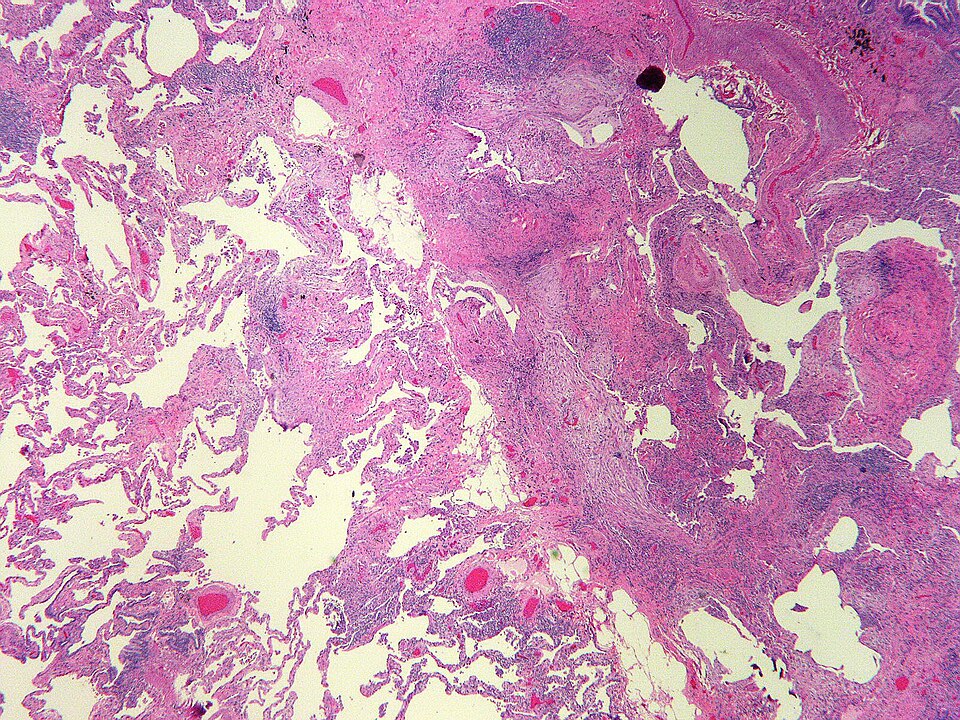
A 58-year-old former shipyard worker presents with progressive dyspnea and a dry cough over 3 years. A CT scan of the chest demonstrates bilateral lower-lobe predominant subpleural honeycombing with traction bronchiectasis. Biopsy of the lung shows the image finding: patchy fibrosis with alternating areas of normal lung parenchyma, fibroblastic foci, and dense collagen deposition. Which of the following best describes the pattern of fibrosis seen on this biopsy?
Yale Rosen, CC BY-SA 2.0 <https://creativecommons.org/licenses/by-sa/2.0>, via Wikimedia Commons
Answer: A. Usual interstitial pneumonia (UIP) is the histologic pattern underlying idiopathic pulmonary fibrosis and is defined by temporal and spatial heterogeneity—areas of dense fibrosis and honeycombing adjacent to normal parenchyma—along with subpleural fibroblastic foci representing the active leading edge of fibrosis. NSIP (option B) shows more temporally uniform, diffuse fibrosis without the patchy heterogeneity and is associated with connective tissue diseases. DIP (option C) shows diffuse macrophage filling of alveoli, most commonly in smokers. Organizing pneumonia (option D) demonstrates intraluminal fibromyxoid plugs within airspaces (Masson bodies), not interstitial collagen deposition. RB-ILD (option E) shows peribronchiolar macrophage accumulation limited to respiratory bronchioles.
Source: Raghu, et al. (2022). Idiopathic Pulmonary Fibrosis (an Update) and Progressive Pulmonary Fibrosis in Adults. American Journal of Respiratory and Critical Care Medicine, 205(9), e18–e47.
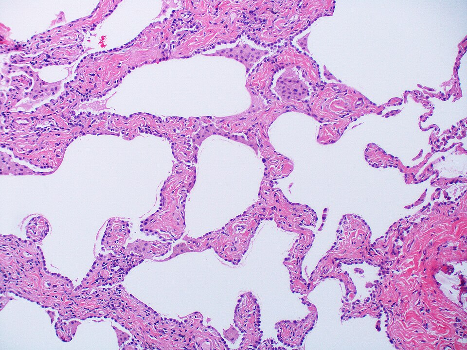
A lung biopsy shows thickened alveolar septa with interstitial fibrosis. Which pulmonary function test finding is most likely present?
Yale Rosen from USA, CC BY-SA 2.0 <https://creativecommons.org/licenses/by-sa/2.0>, via Wikimedia Commons
Answer: C. Interstitial fibrosis thickens the alveolar-capillary membrane, impairing gas exchange and decreasing diffusion capacity. Restrictive lung disease also decreases total lung capacity and lung compliance. Residual volume is not typically increased. FEV1/FVC ratio is often normal or increased in restrictive disease. Therefore, decreased diffusion capacity is the most characteristic finding.
Source: King, et al. (2011). Idiopathic pulmonary fibrosis. The Lancet.
A 45-year-old man presents with progressive exertional dyspnea and dry cough. High-resolution CT scan shows diffuse reticular opacities with honeycombing predominantly in the subpleural and basal regions. Lung biopsy reveals patchy interstitial fibrosis with fibroblastic foci and temporal heterogeneity. Which diagnosis is most likely?
IPFeditor, CC BY-SA 3.0 <https://creativecommons.org/licenses/by-sa/3.0>, via Wikimedia Commons
Answer: D. Idiopathic pulmonary fibrosis (IPF) is characterized by usual interstitial pneumonia (UIP) pattern on histology, including patchy interstitial fibrosis, fibroblastic foci, and temporal heterogeneity. High-resolution CT classically shows basal and subpleural honeycombing with reticular opacities. Sarcoidosis instead shows noncaseating granulomas. Hypersensitivity pneumonitis typically shows poorly formed granulomas and a more bronchiolocentric pattern. Silicosis produces upper-lobe nodular opacities with whorled collagen nodules rather than honeycombing, and ARDS shows diffuse alveolar damage with hyaline membranes.
Source: Raghu, G., et al. (2018). Diagnosis of idiopathic pulmonary fibrosis. American Journal of Respiratory and Critical Care Medicine.
Your next questions are ready.
The question bank picks up from here: it adapts to what you just missed and brings it back right before you'd forget it.
Study smarter with a short weekly email. No spam — unsubscribe anytime.
✓ You're in — talk soon.
Reading is one thing. Remembering is another.
Turn this guide into questions you’ll actually remember
Progress Note turns concepts like this into adaptive questions and spaced review, so difficult topics come back to you exactly when you’re about to forget them.