The abdominal X-ray (KUB) is a quick, low-cost first look at the abdomen, most useful for bowel gas patterns, obstruction, free air, and some calcifications. This guide gives a systematic approach to reading one and explains what it can and cannot reveal.
edit_note
Progress Note Editorial
Evidence-based · For exam prep, not medical advice
update Updated October 2026
Date of last content change
Built by surgery and anesthesiology residents·Written for USMLE Step 1
bolt The 60-second version
check_circleThe abdominal X-ray (KUB) is best for bowel gas patterns, obstruction, and free air.
check_circleRead it systematically: bowel gas, then solid organs, bones, and abnormal densities.
check_circleDilated loops with air-fluid levels suggest bowel obstruction.
check_circleFree air under the diaphragm (on upright films) indicates perforation.
check_circleCT is far more sensitive for most abdominal pathology.
When It Is Used
The abdominal radiograph (often called a KUB, for kidneys-ureters-bladder) is a fast, inexpensive, low-radiation study. It is most useful for evaluating bowel gas patterns and suspected obstruction, detecting free intraperitoneal air (perforation) on an upright or decubitus film, checking the position of tubes and lines, and spotting some radio-opaque stones or foreign bodies. For most detailed abdominal questions, CT has largely replaced it.
A Systematic Approach
Read the film in an organized sequence rather than jumping to an obvious finding. Assess the bowel gas pattern (small bowel is central with valvulae conniventes crossing the full width; large bowel is peripheral with haustra that do not), looking for dilation or an abnormal distribution. Then review the solid organ outlines and the psoas shadows, the bones (spine, pelvis, hips), and any abnormal calcifications or gas outside the bowel.
Key Findings
Several patterns are high-yield. Dilated bowel loops with air-fluid levels on an upright film suggest mechanical obstruction, and the small-versus-large bowel pattern helps localize it; a massively dilated loop can indicate volvulus. Free air under the diaphragm on an upright chest or abdominal film indicates a perforated viscus and is a surgical emergency. Absent or abnormal gas patterns, abnormal calcifications (stones, vascular), and displaced organ outlines are also clues.
Limitations
The abdominal X-ray is insensitive for most pathology — it cannot reliably assess solid organs, appendicitis, early ischemia, or subtle bleeding, and a normal film does not exclude serious disease. When the clinical picture warrants, CT (or ultrasound in specific settings) provides far more information. The KUB is best viewed as a rapid screen for a few specific, high-impact findings rather than a comprehensive study.
Try it now
3 real questions on How to Read an Abdominal X-Ray
Straight from the Progress Note question bank, each with a cited explanation. No account needed.
123
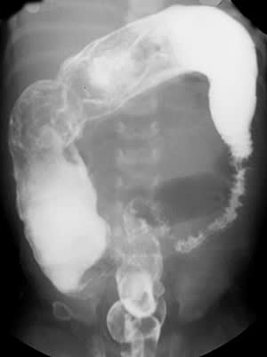
A 2-day-old male newborn has failed to pass meconium within 48 hours of birth and now has abdominal distension and bilious vomiting. A radiograph is shown below. Rectal suction biopsy shows absence of ganglion cells in the submucosal and myenteric plexuses of the narrowed segment, with hypertrophied nerve fibers. The absence of ganglion cells in the affected colonic segment results most directly from which of the following developmental processes?
Copyrightfreeimages, CC0, via Wikimedia Commons
Answer: C. Hirschsprung disease results from failure of neural crest-derived precursor cells to complete their normal craniocaudal migration along the developing gut during early gestation, most commonly due to mutations affecting RET signaling; this leaves the distal-most bowel segment, most often the rectosigmoid colon, permanently devoid of both submucosal and myenteric ganglion cells, producing a tonically contracted, non-relaxing segment with functional obstruction and a radiographic transition zone at the junction with normally innervated, dilated proximal bowel. Because the affected segment never develops normal innervation, this is fundamentally a disorder of neural crest cell migration rather than a postnatal ischemic, autoimmune, myopathic, or purely mechanical process, distinguishing it from each of the incorrect options.
Source: Heuckeroth, R. O. (2018). Hirschsprung disease—Integrating basic science and clinical medicine to improve outcomes. Nature Reviews Gastroenterology & Hepatology, 15(3).
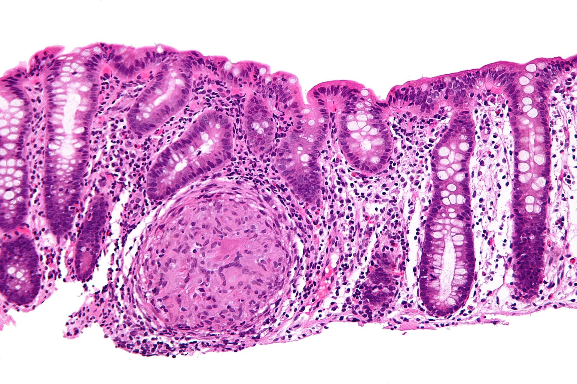
A 26-year-old patient presents with chronic diarrhea, abdominal pain, and weight loss. Colonoscopy shows areas of normal mucosa alternating with deep ulcerations and cobblestoning of the terminal ileum. Which histopathologic finding is most likely?
Nephron, CC BY-SA 3.0 <https://creativecommons.org/licenses/by-sa/3.0>, via Wikimedia Commons
Answer: E. Crohn disease is characterized by transmural inflammation that can affect any part of the GI tract and typically shows skip lesions on gross examination. Noncaseating granulomas may be present on histology and support the diagnosis, though they are not always seen. Ulcerative colitis instead shows continuous mucosal inflammation limited to the colon with crypt abscesses. Celiac disease shows villous atrophy with increased intraepithelial lymphocytes. Pseudomembranes are seen in Clostridioides difficile infection.
Source: Baumgart, D. C., & Sandborn, W. J. (2012). Crohn’s disease. The Lancet.
A 7-year-old boy develops palpable purpura on his lower extremities, abdominal pain, and hematuria following an upper respiratory infection. Skin biopsy shows leukocytoclastic vasculitis with IgA deposition in small vessels. Which underlying mechanism is most likely responsible for this condition?
The original uploader was Okwikikim at English Wikipedia., Public domain, via Wikimedia Commons
Answer: E. IgA vasculitis (Henoch–Schönlein purpura) is caused by immune complex deposition of IgA in small vessels, leading to complement activation and leukocytoclastic vasculitis. It commonly presents after upper respiratory infections and causes palpable purpura, abdominal pain, arthralgias, and renal involvement. Type I hypersensitivity is IgE-mediated and causes allergic reactions rather than vasculitis. Antibody-mediated platelet destruction occurs in ITP, which causes thrombocytopenic purpura without vasculitis. Direct bacterial invasion and granulomatous inflammation do not explain the immune complex deposition pattern.
Source: Piram, M., et al. (2017). IgA vasculitis (Henoch–Schönlein purpura). Joint Bone Spine.
Your next questions are ready.
The question bank picks up from here: it adapts to what you just missed and brings it back right before you'd forget it.
What is an abdominal X-ray (KUB) used for?expand_more
A KUB abdominal X-ray is a fast, low-radiation study best for evaluating bowel gas patterns and obstruction, detecting free air from perforation on upright films, checking tube and line positions, and spotting some radio-opaque stones. For most detailed abdominal problems, CT is used instead.
How do you read an abdominal X-ray systematically?expand_more
Work through it in order: assess the bowel gas pattern (small bowel central with valvulae conniventes, large bowel peripheral with haustra) for dilation or abnormal distribution, then review solid organ outlines and psoas shadows, the bones, and any abnormal calcifications or extraluminal gas.
How does bowel obstruction look on an abdominal X-ray?expand_more
Mechanical bowel obstruction shows dilated bowel loops with air-fluid levels on an upright film, and the small-versus-large bowel gas pattern helps localize it. A massively dilated loop can suggest a volvulus. Distal collapse with proximal dilation supports obstruction.
What does free air on an abdominal X-ray mean?expand_more
Free intraperitoneal air — seen under the diaphragm on an upright chest or abdominal film — indicates a perforated hollow organ (pneumoperitoneum) and is a surgical emergency. Decubitus films can show free air along the flank when a patient cannot sit upright.
Free weekly high-yield email
One high-yield concept + one question, every week
Study smarter with a short weekly email. No spam — unsubscribe anytime.
✓ You're in — talk soon.
Practice makes permanent
Lock this into long-term memory
Progress Note's adaptive engine spaces your reviews automatically — so this stays with you through boards and onto the wards.