Celiac disease is an autoimmune reaction to dietary gluten that damages the small intestinal lining, causing malabsorption in genetically susceptible people. Boards emphasize its varied presentation, the serologic-then-biopsy diagnostic approach, and that treatment is a lifelong gluten-free diet.
Evidence-based · For exam prep, not medical advice
update Updated October 2026
Date of last content change
Built by surgery and anesthesiology residents·Written for USMLE Step 1
bolt The 60-second version
check_circleCeliac disease is an immune reaction to gluten in HLA-DQ2/DQ8–positive individuals.
check_circleIt causes villous atrophy of the small intestine, leading to malabsorption.
check_circlePresentation ranges from diarrhea and weight loss to anemia and dermatitis herpetiformis.
check_circleDiagnosis uses anti-tissue transglutaminase (tTG) IgA, confirmed by duodenal biopsy.
check_circleThe only treatment is a strict, lifelong gluten-free diet.
Pathophysiology
In celiac disease, gliadin peptides from gluten trigger an immune response in genetically predisposed individuals carrying HLA-DQ2 or HLA-DQ8. The reaction damages the small intestinal mucosa, producing villous atrophy, crypt hyperplasia, and intraepithelial lymphocytosis, most prominently in the duodenum. Loss of absorptive surface area causes malabsorption of nutrients, iron, calcium, and fat-soluble vitamins.
Clinical Presentation
The presentation is highly variable. Classic features include chronic diarrhea, steatorrhea, bloating, and weight loss, while many adults present with subtler findings such as iron-deficiency anemia, osteoporosis, or elevated transaminases. In children it can cause failure to thrive. Dermatitis herpetiformis, an intensely itchy vesicular rash, is a specific skin manifestation associated with the disease.
Diagnosis
Testing begins with serology while the patient is still eating gluten: anti-tissue transglutaminase (tTG) IgA is the first-line antibody, with a total IgA level to exclude IgA deficiency. A positive result is confirmed with an upper endoscopy and duodenal biopsy showing villous atrophy. Testing off a gluten-containing diet can produce false negatives, which is a common pitfall.
Management and Complications
The only effective treatment is a strict, lifelong gluten-free diet, which resolves symptoms, heals the mucosa, and normalizes serology over time. Patients are also assessed for nutritional deficiencies and bone density. Untreated or refractory celiac disease carries risks including persistent malabsorption, osteoporosis, and, rarely, enteropathy-associated T-cell lymphoma and small bowel adenocarcinoma.
Try it now
2 real questions on Celiac Disease
Straight from the Progress Note question bank, each with a cited explanation. No account needed.
12
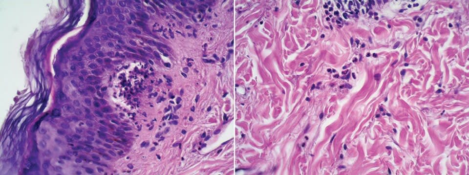
A 28-year-old woman with celiac disease presents with intensely pruritic grouped vesicles and papules on the elbows, knees, and buttocks, arranged in a characteristic clustered pattern. Skin biopsy is shown below. Direct immunofluorescence shows granular IgA deposits specifically at the tips of the dermal papillae. Which of the following best explains the pathophysiology and the relationship between gluten sensitivity, IgA-mediated autoimmunity, and the distinctive immunofluorescence pattern observed in this patient?
Beatriz Di Martino Ortiz, Hugo Macchi, Celeste Valiente Rebull, María Lorena Re Dominguez, Guadalupe Barboza, CC BY 4.0 <https://creativecommons.org/licenses/by/4.0>, via Wikimedia Commons
Answer: B. Dermatitis herpetiformis (DH) is a cutaneous manifestation of celiac disease that presents as intensely pruritic grouped vesicles and papules, typically on extensor surfaces (elbows, knees, buttocks, scalp). The pathophysiology involves a complex interplay between gluten, HLA-DQ2/DQ8 genetics, tissue transglutaminase (tTG), and IgA-mediated autoimmunity. In genetically predisposed individuals, gluten peptides are deamidated by tissue transglutaminase in the intestinal mucosa; these deamidated peptides are presented by HLA-DQ2/DQ8 molecules to CD4+ T cells, triggering Th1 and Th17 responses. Concurrent IgA-secreting B cell responses generate IgA autoantibodies against tissue transglutaminase (intestinal tTG) and epidermal transglutaminase (eTG). These IgA antibodies circulate and deposit in the skin at the dermal-epidermal junction and dermal papillary tips, forming the characteristic granular IgA pattern on direct immunofluorescence. IgA immune complexes at the dermal papillae activate the alternative complement pathway, recruiting neutrophils; neutrophils aggregate and form microabscesses at the dermal-epidermal junction, causing vesicle formation and intense pruritus. Importantly, virtually all patients with DH have concurrent celiac disease on intestinal biopsy (often asymptomatic), establishing the link between gluten ingestion and IgA autoimmunity. Gluten avoidance reduces antigenic stimulation of intestinal and systemic immune responses, resulting in gradual resolution of both intestinal inflammation and skin disease; however, skin lesions resolve more slowly (weeks to months) than GI symptoms due to persistence of skin-resident IgA-secreting plasma cells. Dapsone provides rapid symptom relief by suppressing neutrophil migration but does not address the underlying immunologic process. Option A underestimates IgA's role; option C incorrectly attributes the process to IgE and mast cells; options D and E invoke incorrect mechanisms (collagen/elastin autoimmunity, keratinocyte dysfunction).
Source: Salmi, T. T., et al. (2015). Dermatitis herpetiformis: The newest and the oldest of the autoimmune blistering diseases. Experimental Dermatology, 24(6).
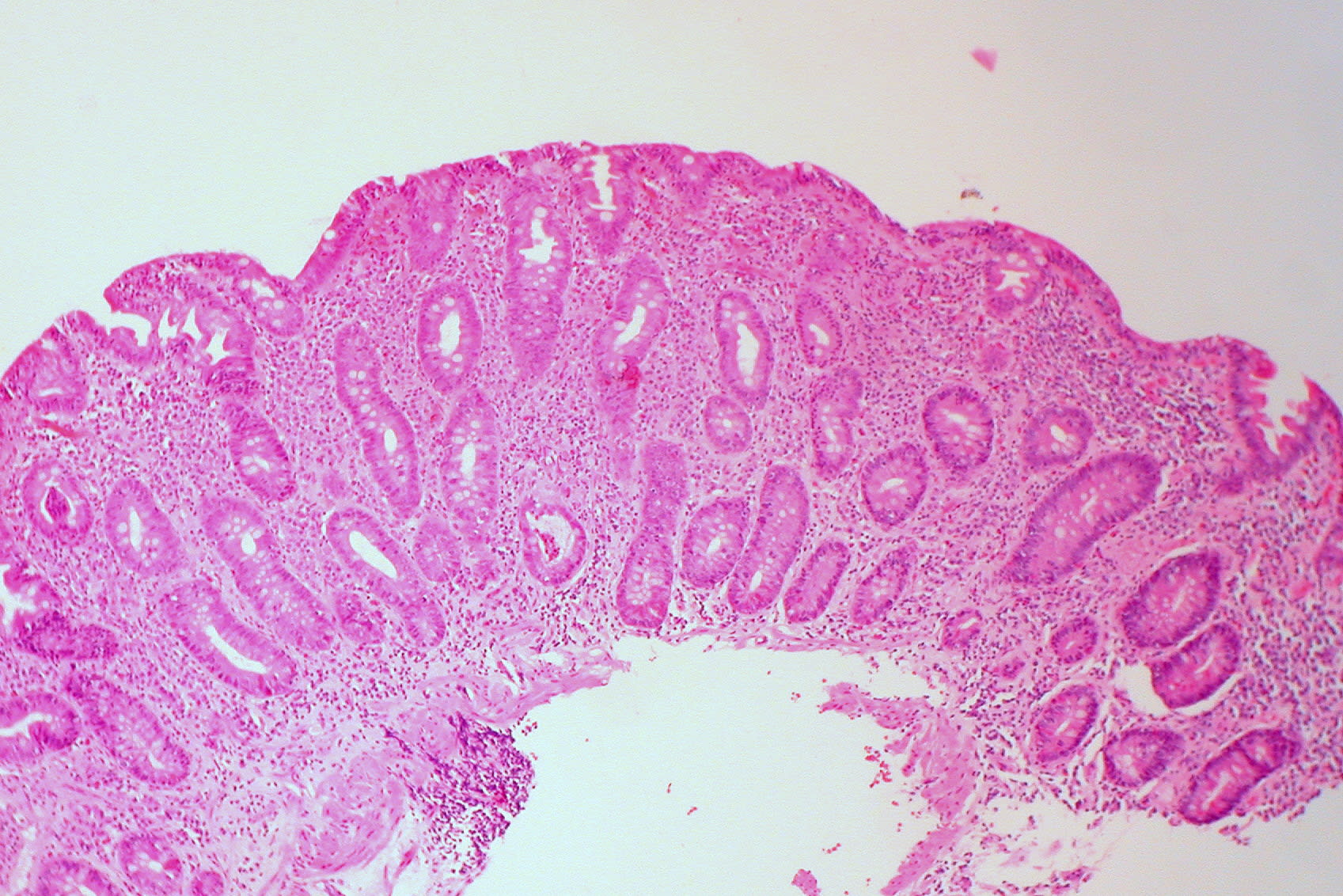
A patient has chronic diarrhea and steatorrhea. Biopsy of small intestine shows villous atrophy and crypt hyperplasia. Which disease is most likely?
Ed Uthman from Houston, TX, USA, CC BY 2.0 <https://creativecommons.org/licenses/by/2.0>, via Wikimedia Commons
Answer: A. Celiac disease is an autoimmune reaction to gluten leading to villous atrophy and crypt hyperplasia in the small intestine. Crohn disease causes transmural inflammation and skip lesions. Ulcerative colitis affects only the colon. IBS has no histologic changes. Pancreatic insufficiency causes malabsorption without villous atrophy.
Source: Lebwohl, et al. (2018). Celiac disease. The Lancet.
Your next questions are ready.
The question bank picks up from here: it adapts to what you just missed and brings it back right before you'd forget it.
Diagnosis starts with serology while the patient is still eating gluten — the anti-tissue transglutaminase (tTG) IgA antibody, along with a total IgA level. A positive result is confirmed with an upper endoscopy and duodenal biopsy showing villous atrophy. Testing off gluten can cause false negatives.
What are the symptoms of celiac disease?expand_more
Symptoms range widely, from chronic diarrhea, bloating, and weight loss to subtler signs such as iron-deficiency anemia, osteoporosis, and fatigue. Children may have failure to thrive, and some patients develop dermatitis herpetiformis, an itchy blistering rash.
What is the treatment for celiac disease?expand_more
The only treatment is a strict, lifelong gluten-free diet, which resolves symptoms, heals the intestinal lining, and normalizes antibody levels. Patients are also evaluated and treated for nutritional deficiencies and low bone density.
Why must you eat gluten before celiac testing?expand_more
Celiac serology and biopsy detect the immune reaction and intestinal damage caused by gluten. If a person has already removed gluten from their diet, the antibodies and villous atrophy may resolve, producing false-negative results, so testing should be done while still consuming gluten.
Free weekly high-yield email
One high-yield concept + one question, every week
Study smarter with a short weekly email. No spam — unsubscribe anytime.
✓ You're in — talk soon.
Reading is one thing. Remembering is another.
Turn this guide into questions you’ll actually remember
Progress Note turns concepts like this into adaptive questions and spaced review, so difficult topics come back to you exactly when you’re about to forget them.