Cirrhosis is the end stage of chronic liver injury: diffuse fibrosis and regenerative nodules replace the normal architecture, throttling blood flow and destroying hepatic function. On USMLE Step 1 it is a rich integration topic — one where a single mechanism, portal hypertension, explains a whole constellation of physical findings. This guide builds cirrhosis from its pathology through portal hypertension to the complications and the Child–Pugh score.
Evidence-based · For exam prep, not medical advice
update Updated October 2026
Date of last content change
Built by surgery and anesthesiology residents
Written for USMLE Step 1 · Free to read
bolt The 60-second version
Cirrhosis is the end stage of chronic liver injury: diffuse fibrosis and regenerative nodules replace the normal architecture, throttling blood flow and destroying hepatic function. On USMLE Step 1 it is a rich integration topic — one where a single mechanism, portal hypertension, explains a whole constellation of physical findings.
check_circleCirrhosis = diffuse fibrosis + regenerative nodules from chronic liver injury, disrupting normal lobular architecture.
check_circleCommon causes: alcohol, chronic hepatitis B and C, and non-alcoholic fatty liver disease (NAFLD).
check_circleFibrosis raises resistance to portal flow → portal hypertension, the source of most complications.
check_circlePortosystemic collaterals: esophageal varices, caput medusae, and internal hemorrhoids; the spleen enlarges and ascites forms.
check_circleLoss of synthetic function lowers albumin and clotting factors (high INR) and raises bilirubin (jaundice).
check_circleThe Child–Pugh score (bilirubin, albumin, INR, ascites, encephalopathy) grades severity as class A, B, or C.
target What you'll be able to do · aligned to USMLE Step 1 · Gastrointestinal System
PATHDescribe the pathology of cirrhosis — fibrosis and regenerative nodules replacing normal lobular architecture — and its major causes.
PATHExplain how increased hepatic resistance produces portal hypertension and its portosystemic collaterals.
DXConnect portal hypertension and hepatic failure to their complications (varices, ascites, encephalopathy, and more).
DXList the components of the Child–Pugh score and what it estimates.
How cirrhosis is tested on Step 1
Step 1 cirrhosis questions usually give a physical finding or a lab pattern and ask for the mechanism. The unifying insight is that two processes — portal hypertension and failing synthetic function — generate nearly every sign. Recognize which of the two is responsible and the answer follows.
workspace_premium The through-line
Two mechanisms explain almost everything: portal hypertension (backpressure) and loss of hepatic function (synthesis and detox).
Pathology and causes
Chronic injury from any cause activates hepatic stellate cells to lay down collagen; surviving hepatocytes regenerate in nodules. The result is a hard, shrunken, nodular liver whose blood flow and function are both compromised.
Architecture. The normal lobule is organized around a central vein with portal triads at its corners; in cirrhosis, regenerative nodules ringed by fibrous bands replace that orderly structure, obstructing flow.
Category
Representative causes
Toxic
Alcohol-related liver disease
Viral
Chronic hepatitis B and C
Metabolic
NAFLD/NASH; hemochromatosis; Wilson disease
Cholestatic/autoimmune
Primary biliary cholangitis; autoimmune hepatitis
Alcohol, chronic viral hepatitis, and NAFLD are the leading causes worldwide.
What makes cirrhosis the common end point of so many diseases is that the liver has only one way to respond to sustained injury: scarring. Whatever the insult — the acetaldehyde of alcohol, the immune attack on hepatitis-infected cells, the lipotoxicity of fatty liver, or iron and copper overload — chronic hepatocyte death recruits Kupffer cells and activates hepatic stellate cells, which transform into collagen-secreting myofibroblasts. Over years, that collagen bridges between portal tracts and central veins, carving the parenchyma into nodules of regenerating hepatocytes with no normal vascular relationships. The two consequences follow directly: blood can no longer flow smoothly through the scarred sinusoids (portal hypertension), and the disorganized, dying hepatocytes can no longer perform the liver’s synthetic and detoxifying work (hepatic insufficiency). Importantly, early fibrosis can regress if the cause is removed, but established cirrhosis with its nodular architecture is largely irreversible.
Portal hypertension and its collaterals
The fibrotic liver resists the portal blood trying to pass through it, so pressure rises in the portal system. Blood is forced backward into portosystemic anastomoses — the collaterals that produce cirrhosis’s classic signs.
Portal hypertension. Increased hepatic resistance backs blood into collateral veins: esophageal varices, caput medusae, and internal hemorrhoids, while the spleen enlarges, ascites accumulates, and gut toxins bypass the liver to cause encephalopathy.
Each collateral is a portosystemic anastomosis — a point where the portal circulation meets the systemic veins. When portal pressure climbs, these normally trivial connections dilate to decompress the system. At the lower esophagus, the left gastric vein anastomoses with the azygos system to form esophageal varices, which can bleed catastrophically. At the umbilicus, recanalized paraumbilical veins produce the dilated abdominal wall veins of caput medusae. In the rectum, the superior rectal vein connects with the middle and inferior rectal veins to form internal hemorrhoids. Meanwhile blood dams back into the splenic vein, enlarging the spleen, and the combination of high sinusoidal pressure and low oncotic pressure from hypoalbuminemia drives fluid into the peritoneum as ascites.
Complications
Beyond the collaterals, cirrhosis produces a set of high-yield complications from portal hypertension and hepatic failure together.
Ammonia-lowering therapy (lactulose) treats encephalopathy; spontaneous bacterial peritonitis is a feared infection of ascitic fluid.
Two of these deserve special attention because they are both common and deadly. Spontaneous bacterial peritonitis is infection of pre-existing ascitic fluid without an obvious source; it is diagnosed by a raised neutrophil count in the ascites and is a frequent, treatable cause of decompensation in a previously stable cirrhotic. Hepatorenal syndrome is a functional kidney failure driven by intense splanchnic vasodilation that steals blood flow from the kidneys — the kidneys themselves are structurally normal, which is why the injury can reverse if liver function is restored. Recognizing both as consequences of the same portal-hypertensive physiology, rather than as separate organ diseases, is exactly the kind of integration Step 1 rewards.
error Common trap
Hepatic encephalopathy is precipitated by anything that raises ammonia or nitrogen load — GI bleeding, infection, constipation, or dehydration — which is why treatment targets ammonia with lactulose.
Grading severity: Child–Pugh
Severity and prognosis are estimated by the Child–Pugh score, a favorite because its five components map onto the two core mechanisms.
Child–Pugh components. Bilirubin, albumin, and INR reflect synthetic/excretory function; ascites and encephalopathy reflect portal hypertension and hepatic failure. Together they grade class A, B, or C.
functions Worked example — localize the sign
A patient with alcohol-related cirrhosis develops dilated veins radiating from the umbilicus across the abdominal wall. What is this sign and what causes it?
Dilated abdominal-wall veins around the umbilicus is the physical finding.
The mechanism is portal hypertension forcing blood into paraumbilical collaterals.
These connect the portal system to systemic veins of the abdominal wall.
Answer. Caput medusae — a portosystemic collateral of portal hypertension, in which recanalized paraumbilical veins carry high-pressure portal blood into the systemic circulation of the abdominal wall.
How to study it — and the traps to avoid
Cirrhosis rewards mapping every sign back to portal hypertension or lost hepatic function.
Fix the pathology first: fibrosis + regenerative nodules replacing normal architecture.
Trace portal hypertension to its collaterals (varices, caput medusae, hemorrhoids) and to splenomegaly and ascites.
Attribute low albumin, high INR, and jaundice to failed synthetic/excretory function.
Know the Child–Pugh components and that lactulose treats encephalopathy by lowering ammonia.
workspace_premium Integration
Coagulopathy connects to the hematology workup, and the hemodynamics of portal hypertension parallel the pressure concepts in cardiovascular physiology.
Straight from the Progress Note question bank, each with a cited explanation. No account needed.
123
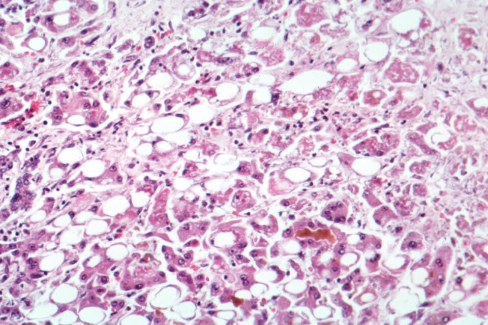
A 52-year-old man with a 15-year history of alcohol use disorder presents with jaundice, abdominal distension, and melena. Laboratory studies reveal elevated transaminases (AST 320 U/L, ALT 180 U/L), elevated bilirubin (5.2 mg/dL), coagulopathy (INR 2.8), and evidence of portal hypertension. Liver biopsy is shown below. Which of the following best explains the pathophysiology of alcoholic hepatitis and how the progression from steatosis through steatohepatitis to cirrhosis relates to the specific histologic findings observed in this patient?
Countincr at English Wikipedia., CC BY-SA 2.5 <https://creativecommons.org/licenses/by-sa/2.5>, via Wikimedia Commons.
Answer: B. Alcoholic hepatitis results from a multi-hit mechanism: ethanol metabolism by ADH and CYP2E1 generates acetaldehyde and oxidative stress (CYP2E1-derived reactive oxygen species). Acetaldehyde forms stable protein adducts (malondialdehyde-acetaldehyde, or MAA adducts) that are recognized as neo-antigens and trigger both innate immune activation (TLR-mediated inflammatory cytokine release) and adaptive immune responses (CD8+ T-cell infiltration). Simultaneously, oxidative stress damages cellular proteins and lipids, impairing mitochondrial function and triggering hepatocyte apoptosis and necrosis. Mallory-Denk bodies are aggregates of hyperphosphorylated proteins (ubiquitin, heat shock proteins) that accumulate in hepatocytes damaged by oxidative stress; their presence indicates severe hepatocellular injury. The neutrophilic infiltrate seen in alcoholic steatohepatitis reflects both innate immune activation and recruitment by chemokines released from damaged hepatocytes. Fibrosis results from stellate cell activation by inflammatory cytokines (TNF-alpha, TGF-beta) released from dying hepatocytes and activated Kupffer cells. Option A incorrectly denies the importance of inflammation in steatosis progression; options C, D, and E invoke incorrect mechanisms (stellate cell inhibition by alcohol, viral etiology, apoptosis without immune contribution).
Source: Gao, B., & Bataller, R. (2011). Alcoholic liver disease: Pathogenesis and new therapeutic targets. Gastroenterology, 141(5).
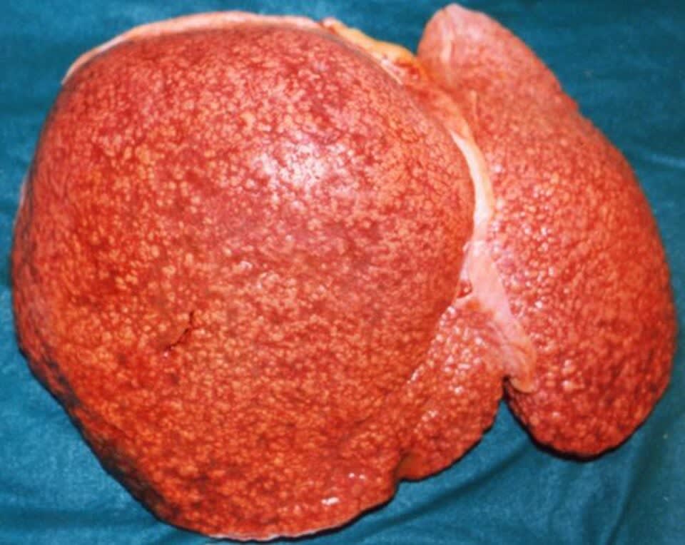
A 58-year-old man with a 30-year history of alcohol use disorder undergoes liver transplantation for end-stage liver disease. Gross pathology specimen is shown below. Histology demonstrates extensive bridging fibrosis with thin fibrous bands connecting portal tracts to central veins and separating regenerative hepatocyte nodules. Which of the following best explains how chronic ethanol-induced hepatocyte injury, stellate cell activation, and aberrant extracellular matrix remodeling collectively lead to the morphologic findings observed?
Amadalvarez, CC BY-SA 4.0 <https://creativecommons.org/licenses/by-sa/4.0>, via Wikimedia Commons
Answer: D. Alcoholic cirrhosis results from a chronic necrosis-regeneration-fibrosis cycle: ethanol-derived acetaldehyde and oxidative stress (via CYP2E1 metabolism) damage hepatocytes, triggering apoptosis and necroinflammation; dying hepatocytes release cytokines (TGF-beta, PDGF, TNF-alpha) that activate hepatic stellate cells toward a myofibroblast phenotype; myofibroblasts produce excessive collagen and deposit extracellular matrix. Simultaneously, surviving hepatocytes proliferate to regenerate lost liver mass, but in the setting of continuous injury, regeneration becomes dysfunctional and combines with progressive fibrosis to distort normal hepatic architecture—bridging fibrosis obliterates the normal zone-based hepatic structure. Option A oversimplifies the mechanism by attributing fibrosis solely to hepatocyte death; option B emphasizes steatosis as a necessary precursor (steatosis alone does not cause cirrhosis); option C incorrectly suggests ethanol inhibits stellate cell activation; option E denies the role of regeneration and architectural distortion in cirrhosis formation.
Source: Singal, A. K., & Mathurin, P. (2021). Diagnosis and treatment of alcohol-associated liver disease: A review. JAMA, 326(2).
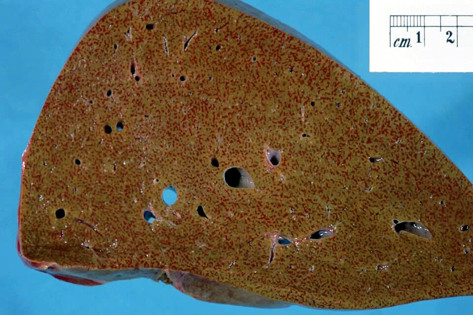
What is the most likely diagnosis?
Narraburra, CC0, via Wikimedia Commons
Answer: B. Chronic passive congestion from right-sided heart failure is the classic cause of the 'nutmeg liver' pattern, in which elevated central venous pressure is transmitted to the hepatic sinusoids, producing centrilobular sinusoidal dilation, congestion, and hepatocyte atrophy that alternates with less affected periportal tissue. Micronodular cirrhosis (A) shows diffuse small regenerative nodules separated by fibrous septa rather than a reticular congestion pattern. Budd-Chiari syndrome (C) can produce a grossly similar centrilobular congestion pattern from hepatic venous outflow obstruction, but it typically causes more acute, severe centrilobular hemorrhagic necrosis with prominent ascites and is usually associated with an underlying hypercoagulable state rather than systemic venous congestion from cardiac failure. Hereditary hemochromatosis (D) produces a diffusely brown, iron-laden liver from hemosiderin deposition rather than a mottled congestive pattern. Nonalcoholic fatty liver disease (E) produces a diffusely yellow, greasy-appearing liver from macrovesicular steatosis, without the dark centrilobular congestion seen here.
Source: Samsky, M. D., et al. (2013). Cardiohepatic interactions in heart failure: An overview and clinical implications. Journal of the American College of Cardiology.
Your next questions are ready.
The question bank picks up from here: it adapts to what you just missed and brings it back right before you'd forget it.
What are the most common causes of cirrhosis?expand_more
The most common causes are alcohol-related liver disease, non-alcoholic steatohepatitis (NASH), and chronic hepatitis B and C infection. Less common causes include autoimmune hepatitis, hemochromatosis, Wilson disease, alpha-1-antitrypsin deficiency, and primary biliary or sclerosing cholangitis.
What is spontaneous bacterial peritonitis?expand_more
Spontaneous bacterial peritonitis (SBP) is infection of ascitic fluid without an obvious source, occurring in patients with cirrhosis and ascites. It is diagnosed when the ascitic fluid absolute neutrophil count exceeds 250 cells per microliter and is treated empirically with a third-generation cephalosporin such as cefotaxime.
How is hepatic encephalopathy treated?expand_more
Hepatic encephalopathy is treated with lactulose, which traps ammonia in the gut for excretion, and rifaximin, a poorly absorbed antibiotic that reduces ammonia-producing bacteria. Identifying and correcting precipitants such as GI bleeding, infection, constipation, and electrolyte disturbances is equally important.
Why does cirrhosis increase the risk of liver cancer?expand_more
Chronic injury and the continuous cycle of hepatocyte death and regeneration in cirrhosis promote genetic mutations that can lead to hepatocellular carcinoma. Because of this risk, patients with cirrhosis undergo periodic surveillance, typically with liver ultrasound every six months.
Study smarter with a short weekly email. No spam — unsubscribe anytime.
✓ You're in — talk soon.
Reading is one thing. Remembering is another.
Turn this guide into questions you’ll actually remember
Progress Note turns concepts like this into adaptive questions and spaced review, so difficult topics come back to you exactly when you’re about to forget them.