How pericardial disease is tested on Step 1
Vignettes describe chest pain that changes with position, a hypotensive patient with distended neck veins after trauma or malignancy, or chronic right-sided congestion after radiation or TB.
A slowly growing effusion can reach a liter without symptoms because the pericardium stretches. A rapid 150 mL of blood (trauma, ventricular rupture) can cause tamponade.
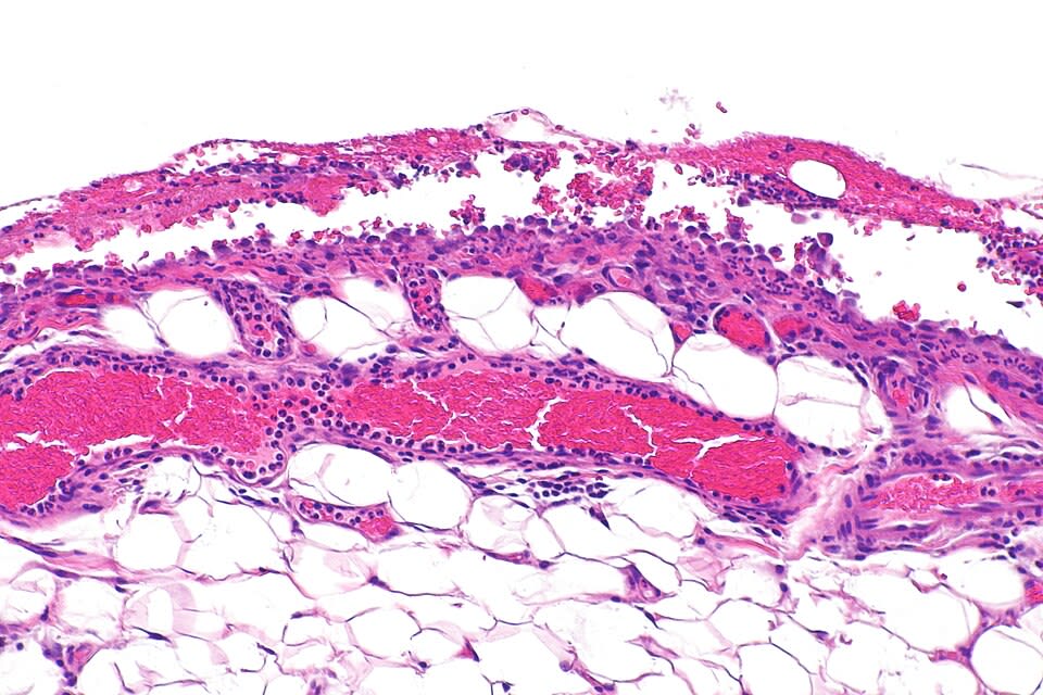
Acute pericarditis
Inflammation of the pericardial layers causes pain and a characteristic ECG.
| Feature | Pericarditis | STEMI |
|---|---|---|
| Pain | Sharp, pleuritic, better sitting forward | Pressure, not positional |
| ST elevation | Diffuse, concave ("saddle-shaped") | Regional, convex, with reciprocal depression |
| PR segment | Depressed (elevated in aVR) | Usually normal |
| Q waves | Absent | May develop |
| Exam | Friction rub (up to three components) | May have S4, new murmur |
Treatment: NSAIDs (or aspirin after MI) plus colchicine for 3 months; avoid NSAIDs other than aspirin early after MI.
Cardiac tamponade
Fluid under pressure compresses all four chambers, limiting diastolic filling. Stroke volume and blood pressure fall.
| Finding | Explanation |
|---|---|
| Beck triad: hypotension, JVD, muffled heart sounds | Low output, high venous pressure, fluid around the heart |
| Pulsus paradoxus > 10 mm Hg | Ventricular interdependence |
| Electrical alternans | The heart swings within the fluid |
| Low-voltage QRS | Fluid damps the signal |
| Echo: RA and RV diastolic collapse | Pericardial pressure exceeds chamber pressure |
Treatment is urgent pericardiocentesis; give IV fluids and avoid diuretics, vasodilators, and positive-pressure ventilation if possible.
Constrictive pericarditis
Chronic inflammation leaves a thick, fibrotic, often calcified pericardium that stops the heart from filling in late diastole.
Clinical application
Trauma and malignancy are classic tamponade settings.
A man with a stab wound to the left chest has blood pressure 78/50, distended neck veins, distant heart sounds, and a systolic pressure that drops 18 mm Hg on inspiration. Lungs are clear bilaterally and the trachea is midline. What is the diagnosis and treatment?
- Hypotension, JVD, and muffled heart sounds form Beck triad.
- A fall of more than 10 mm Hg on inspiration is pulsus paradoxus.
- Clear breath sounds and a midline trachea argue against tension pneumothorax, the main alternative.
- Rapid accumulation of blood in the pericardium explains tamponade from a small volume.
Answer. Traumatic cardiac tamponade. Treat with immediate pericardiocentesis or surgical drainage; IV fluids temporarily support preload.