Ventricular fibrillation (VF) is a chaotic, disorganized ventricular rhythm that produces no effective cardiac output, causing sudden cardiac arrest. Boards emphasize recognizing it as a shockable rhythm, its common causes, and the immediate response of CPR and defibrillation.
Evidence-based · For exam prep, not medical advice
update Updated October 2026
Date of last content change
Built by surgery and anesthesiology residents·Written for USMLE Step 1
bolt The 60-second version
check_circleVentricular fibrillation is disorganized electrical activity with no effective contraction.
check_circleIt causes sudden cardiac arrest — no pulse, no output.
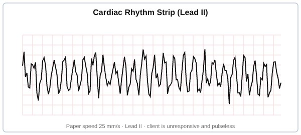
check_circleThe ECG shows chaotic, irregular waveforms without identifiable QRS complexes.
check_circleIt is a shockable rhythm: immediate defibrillation is the definitive treatment.
check_circleHigh-quality CPR and early defibrillation drive survival.
What It Is
Ventricular fibrillation is a life-threatening arrhythmia in which the ventricles quiver chaotically instead of contracting in a coordinated way, so the heart pumps no blood. It is one of the main rhythms causing sudden cardiac arrest. Without immediate treatment, the absence of cardiac output leads to loss of consciousness within seconds and death within minutes.
Causes
The most common precipitant is acute myocardial ischemia or infarction, which destabilizes the ventricular myocardium. Other causes include underlying structural heart disease and cardiomyopathies, electrolyte abnormalities (such as potassium and magnesium disturbances), prolonged QT and certain drugs, and channelopathies. Ventricular tachycardia can degenerate into ventricular fibrillation.
Recognition
On the monitor or ECG, ventricular fibrillation appears as a chaotic, irregular, wavering baseline without discernible P waves, QRS complexes, or T waves; it may be coarse or fine. Clinically, the patient is pulseless and unresponsive. Recognizing a pulseless patient with this pattern as a shockable rhythm is the critical first step, and it must be distinguished from artifact and from asystole.
Treatment
Ventricular fibrillation is treated with immediate defibrillation — an unsynchronized electrical shock — which is the only definitive therapy, combined with high-quality CPR to maintain perfusion between shocks. In the ACLS algorithm, CPR and early defibrillation are prioritized, with epinephrine and antiarrhythmics (amiodarone or lidocaine) as adjuncts, while reversible causes are sought and corrected. After successful resuscitation, an implantable defibrillator is often considered.
Try it now
3 real questions on Ventricular Fibrillation
Straight from the Progress Note question bank, each with a cited explanation. No account needed.
123
From our NCLEX bank
A nurse observes the rhythm shown on the monitor of a client who is suddenly unresponsive and pulseless. How should the nurse interpret and respond to this rhythm?
Answer: B. The strip shows chaotic, disorganized electrical activity with no identifiable QRS complexes, which is ventricular fibrillation; in a pulseless, unresponsive client this is a cardiac emergency requiring immediate CPR and defibrillation (option B). This is not sinus tachycardia (A), which has organized complexes, and it is clearly not normal sinus rhythm (C). Atrial fibrillation (D) is an irregular rhythm with a pulse and does not cause this pulseless chaotic pattern. The nurse starts high-quality chest compressions, calls for help and the defibrillator, and defibrillates as soon as it is available. Early defibrillation is the key to survival in ventricular fibrillation.
Source: Harding, M. M., Kwong, J., Hagler, D., & Reinisch, C. (2023). Lewis's medical-surgical nursing (12th ed.). Elsevier.
From our NCLEX bank
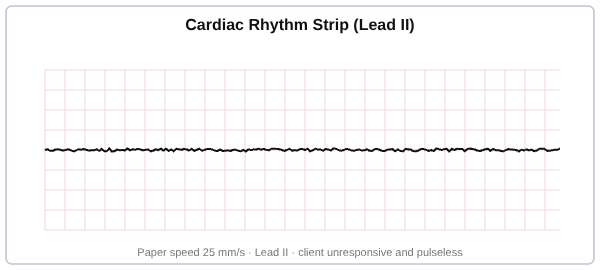
A nurse observes the rhythm shown on the monitor of a client who is unresponsive and pulseless. How should the nurse interpret and respond to this rhythm?
Answer: A. The strip shows a flat line with no electrical activity, which is asystole; in a pulseless client the nurse confirms it in a second lead, begins CPR, and does not defibrillate because asystole is a non-shockable rhythm (option A). Ventricular fibrillation (B) shows chaotic waveforms and is shockable, which does not match a flat line. This is clearly not normal sinus rhythm (C). Sinus bradycardia (D) has organized complexes with a pulse and does not apply to a pulseless, flatline rhythm. The nurse provides high-quality CPR, gives epinephrine per protocol, and searches for reversible causes. Confirming asystole in another lead rules out a disconnected lead.
Source: Harding, M. M., Kwong, J., Hagler, D., & Reinisch, C. (2023). Lewis's medical-surgical nursing (12th ed.). Elsevier.
From our NCLEX bank
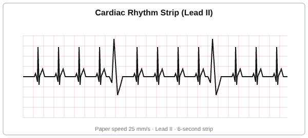
A nurse observes the rhythm strip shown on the monitor of a client who has a palpable pulse and is asymptomatic. How should the nurse interpret and respond to this rhythm?
Answer: C. The strip shows underlying sinus beats interrupted by early, wide, bizarre complexes, which are premature ventricular contractions (PVCs); the nurse assesses the client and checks electrolytes such as potassium and magnesium, because imbalances are a common cause (option C). This is not normal sinus rhythm (A) because of the ectopic wide beats. Ventricular fibrillation (B) is a chaotic rhythm with no organized complexes in a pulseless client, which does not match this strip. Atrial fibrillation (D) is an irregularly irregular rhythm without discrete P waves and does not produce isolated wide premature beats. The nurse monitors the frequency of PVCs, since increasing or multifocal PVCs can precede more dangerous dysrhythmias.
Source: Harding, M. M., Kwong, J., Hagler, D., & Reinisch, C. (2023). Lewis's medical-surgical nursing (12th ed.). Elsevier.
Your next questions are ready.
The question bank picks up from here: it adapts to what you just missed and brings it back right before you'd forget it.
Ventricular fibrillation is a chaotic, disorganized ventricular rhythm in which the ventricles quiver rather than contract, so the heart pumps no blood. It causes sudden cardiac arrest and is fatal within minutes without immediate treatment.
How is ventricular fibrillation treated?expand_more
Ventricular fibrillation is treated with immediate defibrillation (an unsynchronized shock), the only definitive therapy, together with high-quality CPR between shocks. Epinephrine and antiarrhythmics like amiodarone are used as adjuncts, and reversible causes are corrected.
What does ventricular fibrillation look like on an ECG?expand_more
On ECG, ventricular fibrillation appears as a chaotic, irregular, wavering baseline with no identifiable P waves, QRS complexes, or T waves. It may be coarse or fine, and the patient is pulseless and unresponsive.
What causes ventricular fibrillation?expand_more
The most common cause is acute myocardial ischemia or infarction. Other causes include structural heart disease and cardiomyopathies, electrolyte abnormalities, prolonged QT and certain drugs, and channelopathies. Ventricular tachycardia can degenerate into ventricular fibrillation.
Free weekly high-yield email
One high-yield concept + one question, every week
Study smarter with a short weekly email. No spam — unsubscribe anytime.
✓ You're in — talk soon.
Reading is one thing. Remembering is another.
Turn this guide into questions you’ll actually remember
Progress Note turns concepts like this into adaptive questions and spaced review, so difficult topics come back to you exactly when you’re about to forget them.