The Cornell note-taking method is a structured system that splits the page into a cue column, a notes area, and a summary, turning passive note-taking into active review. It is popular with students because it builds recall and organization into the notes themselves.
edit_note
Progress Note Editorial
Evidence-based · For exam prep, not medical advice
update Updated October 2026
Date of last content change
Built by surgery and anesthesiology residents·Written for USMLE Step 1
bolt The 60-second version
check_circleThe page is divided into a narrow cue column, a wide notes area, and a bottom summary.
check_circleNotes go in the main area during class or reading.
check_circleQuestions and keywords go in the cue column afterward.
check_circleA short summary at the bottom consolidates the page.
check_circleThe cue column enables self-testing by covering the notes.
The Layout
The Cornell system divides each page into three zones: a wide right-hand notes column (about two-thirds of the page), a narrow left-hand cue column, and a summary bar across the bottom. This simple structure is the entire method, and it forces you to process information at three moments — while recording, while reviewing, and while summarizing — rather than only once.
During Class or Reading
While listening or reading, capture the main ideas, facts, and examples in the large notes column using concise phrases rather than full sentences. Do not try to fill the cue column yet; the goal is to record the substance efficiently. Leaving the cue column and summary blank at this stage is intentional, because they are completed during active review.
After Class: Cues and Summary
Soon after, distill the notes into questions and keywords in the cue column — each one a prompt for a chunk of the notes beside it. Then write a one- to three-sentence summary of the whole page at the bottom in your own words. This after-class pass is where most of the learning happens, because it requires you to reorganize and condense the material.
Reviewing With Cornell Notes
The layout is built for self-testing. Cover the notes column and use the cue-column questions to recall the material from memory, checking yourself against the notes — a form of active recall. Reading the bottom summaries lets you review many pages quickly. Combining Cornell notes with spaced repetition turns a set of notes into an efficient study tool.
Try it now
Try 3 real questions
Straight from the Progress Note question bank, each with a cited explanation. No account needed.
123
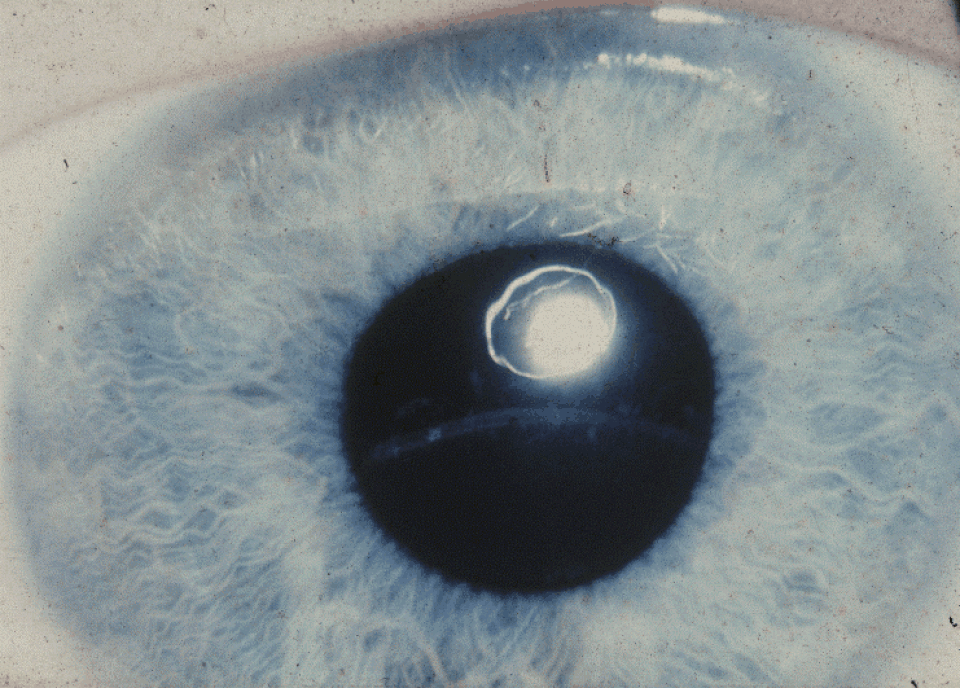
A tall, thin 10-year-old boy has intellectual disability and a history of an unprovoked deep vein thrombosis. A photo of his slit-lamp exam is shown below. Urine nitroprusside testing is positive, and plasma amino acid analysis shows markedly elevated homocysteine and methionine. Which enzyme deficiency and mechanism best account for both the connective tissue and thrombotic findings?
National Eye Institute, Public domain, via Wikimedia Commons
Answer: A. Classic homocystinuria results from cystathionine beta-synthase deficiency, which blocks the transsulfuration pathway and causes accumulation of both homocysteine and its precursor methionine; elevated homocysteine promotes endothelial damage and a prothrombotic state (explaining the thromboembolic risk) and also interferes with normal lysine-derived collagen and elastin cross-linking, contributing to the connective tissue laxity responsible for lens dislocation (characteristically downward, helping distinguish it clinically from the upward lens dislocation typical of Marfan syndrome) and skeletal features. Option B incorrectly proposes MTHFR deficiency, which causes homocystinuria with low (not elevated) methionine due to impaired remethylation, the opposite pattern from what is described. Option C incorrectly attributes the findings to a fibrillin-1 structural defect, the mechanism underlying Marfan syndrome, a distinct condition with a different lens dislocation direction and no thrombotic predisposition. Option D incorrectly dismisses the enzymatic block, attributing methionine elevation to diet alone. Option E incorrectly substitutes an unrelated primary platelet disorder for the amino acid metabolism defect actually responsible.
Source: Morris, A. A., et al. (2017). Guidelines for the diagnosis and management of cystathionine beta-synthase deficiency. Journal of Inherited Metabolic Disease.
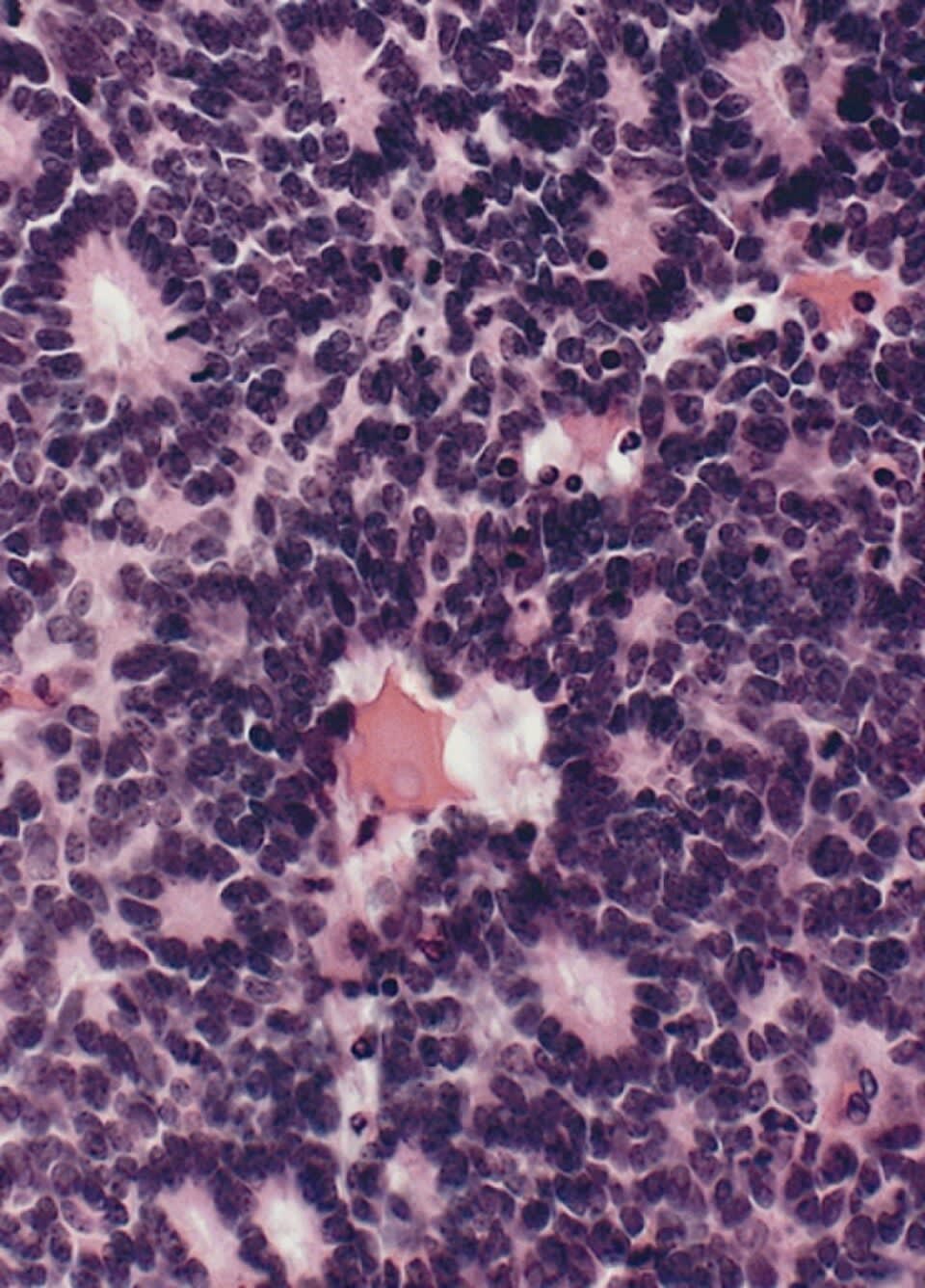
What is the diagnosis?
The Armed Forces Institute of Pathology (AFIP), Public domain, via Wikimedia Commons
Answer: E. Retinoblastoma, the most common intraocular malignancy of childhood, arises from biallelic loss of the RB1 tumor suppressor gene and classically presents with leukocoria. Histology shows small round blue cells forming Flexner-Wintersteiner rosettes, distinguished by a true central lumen, unlike the Homer-Wright rosettes (cells encircling a tangle of neural processes without a lumen) seen in neuroblastoma and medulloblastoma. Ewing sarcoma shows sheets of small round blue cells without rosette formation, and astrocytic hamartomas show bland, glial-appearing cells rather than malignant small round blue cells.
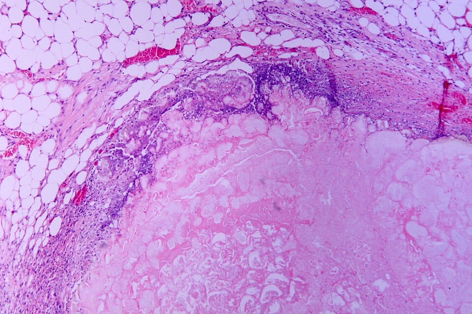
A patient presents with severe epigastric pain radiating to the back. Serum lipase is elevated. Histology shows fat necrosis with calcium soap formation. Which process is responsible?
Patho, CC BY-SA 3.0 <https://creativecommons.org/licenses/by-sa/3.0>, via Wikimedia Commons
Answer: E. Acute pancreatitis is caused by premature activation of pancreatic enzymes, especially trypsin, leading to autodigestion of pancreatic tissue. Lipase release causes fat necrosis and saponification due to calcium binding. Bile reflux is a contributing factor but not the primary mechanism. Viral infection is not typical. Autoimmune pancreatitis is a distinct chronic condition.
Source: Forsmark, et al. (2016). Acute pancreatitis. New England Journal of Medicine.
Your next questions are ready.
The question bank picks up from here: it adapts to what you just missed and brings it back right before you'd forget it.
What is the Cornell note-taking method?expand_more
The Cornell method divides each page into a wide notes column, a narrow left-hand cue column for questions and keywords, and a summary at the bottom. You take notes during class, add cues and a summary afterward, and use the cue column to self-test, building review into the notes themselves.
How do you use Cornell notes to study?expand_more
Cover the notes column and use the questions in the cue column to recall the material from memory, then check yourself against the notes — a form of active recall. Reading the bottom summaries lets you review many pages quickly, and pairing this with spaced repetition improves retention.
Why is the Cornell method effective?expand_more
It is effective because it forces you to process material at three points — recording notes, creating cues, and summarizing — rather than passively once. The cue column also builds in self-testing, which is far more effective for memory than rereading.
When do you fill in the cue column?expand_more
The cue column is filled in after class or reading, not during. During the session you record notes in the main column; afterward you distill them into questions and keywords in the cue column and write a summary, which is where most of the learning happens.
Free weekly high-yield email
One high-yield concept + one question, every week
Study smarter with a short weekly email. No spam — unsubscribe anytime.
✓ You're in — talk soon.
Practice makes permanent
Lock this into long-term memory
Progress Note's adaptive engine spaces your reviews automatically — so this stays with you through boards and onto the wards.